Anatomy Ultrasound
The 20 week ultrasound or anatomy scan is an eagerly anticipated ultrasound for parents. Some wavelengths are intended to break up tissue but the wavelengths that we use for diagnostics imaging are not at all in the ranges that could cause significant tissue damage.
 Anatomy Ultrasound Maternal Fetal Associates Of The Mid
This is a detailed scan of your babybabies anatomy.

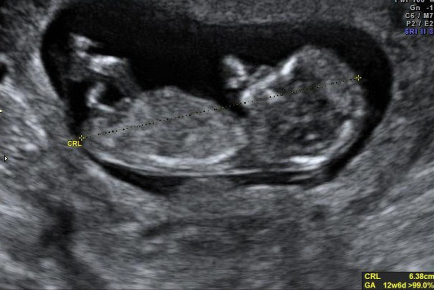
Anatomy ultrasound. There are different wavelengths of ultrasounds. There are two main types of fetal ultrasound exams. The second trimester extends from 13 weeks and 0 days to 27 weeks and 6 days of gestation although the majority of these studies are performed between 18 and 23 weeks.
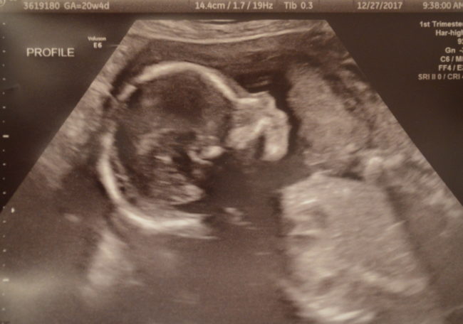
Luckily all was well. Find out what youll see when you have yours. Most anatomy scans are performed in the second trimester of pregnancy typically at 20 weeks but they can be done anytime between 18 weeks and 22 weeks.
It became hugely important for me to have three sets of names going into the ultrasound so that we were. Skull shape integrity bpd and hc measurements. In women at high risk for preterm delivery multiple pregnancies previous preterm birth abnormalities of the uterus or previous cervical surgery we may also carry out a transvaginal scan to measure the length of the cervix.
Those who want to can find out the sex of the baby if desired. The following fetal parts are checked during the anatomy ultrasound. The anatomy scan is a level 2 ultrasound which is typically performed on pregnant women between 18 and 22 weeks.
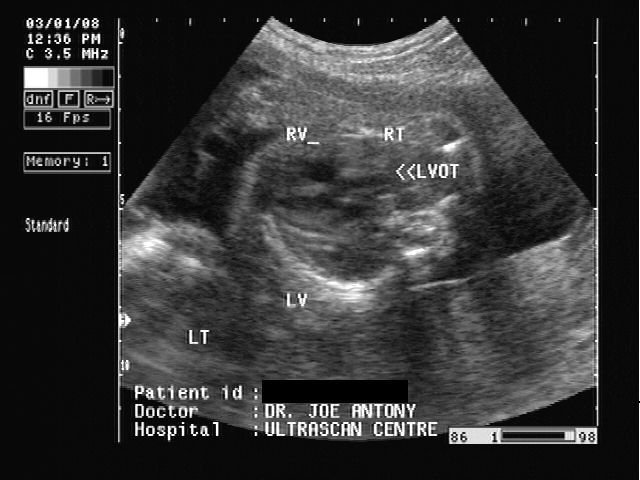
Heart rate rhythm 4 chamber views. Ultrasound means outside of the range of human hearing. I have a brother with downs syndrome trisomy 21 though and i told my sonographer.
I was planning a home birth and felt that it was important to ensure there were no serious risk factors present so that i could switch my birth to the hospital if needed. A transabdominal fetal ultrasound is done by moving a transducer. If you have a condition that needs to be monitored such as carrying multiples you may have more than one detailed ultrasound.
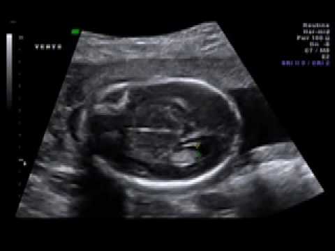
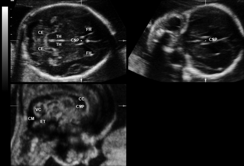
Brain ventricles choroid plexus mid brain posterior fossa cerebellum cisterna magna. With this type of fetal ultrasound a wandlike device called. The second trimester scan is a routine ultrasound examination in many countries that is primarily used to assess fetal anatomy and detect the presence of any fetal anomalies.
As the anatomy scan approached i began to build the what if scenarios up in my head. The anatomy ultrasound was the only one i had. When a level 2 ultrasound is done.
The scan is performed transabdominally. Neck nuchal fold thickness.
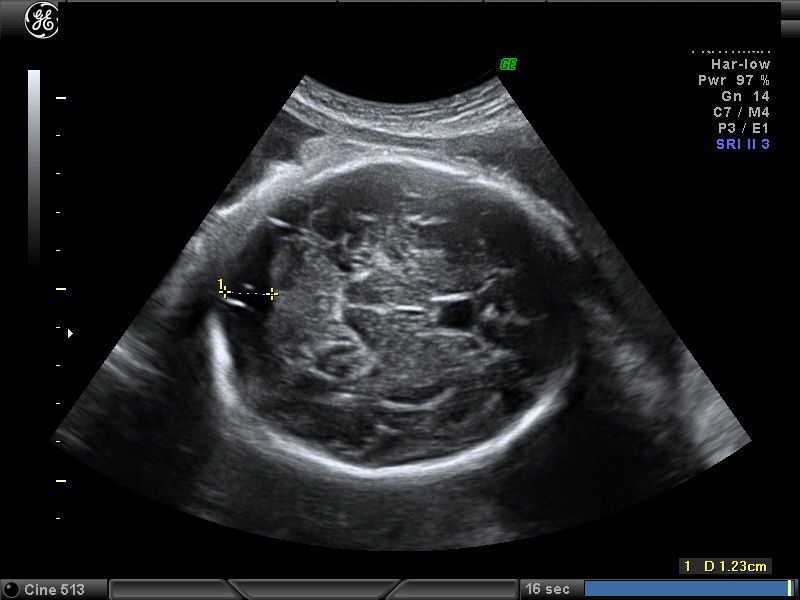 Ultrasound Images Of Fetal Brain
 Ultrasound Assessment Of Normal Fetal Anatomy Chapter 5
 An Ultrasound For My Birthday Stories Thyme
 Scrotal Ultrasound Startradiology
 A Gallery Of High Resolution Ultrasound Color Doppler 3d
 Ultrasound Anatomy Scan 19 Weeks Musc Health
 Ultrasound 3 Anatomy Scan Zygotta
 19 Weeks Our Anatomy Ultrasound The Love Notes Blog
 18 Week 3 Day Anatomy Ultrasound Chihuahuaesque Flickr
 The Radiology Assistant Normal Values Ultrasound
 Free Chapter Normal Cns Ultrasound Brain Anatomy Ob Images
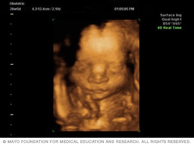 Fetal Ultrasound Mayo Clinic
 Gender Anatomy Development Ultrasound 20 Wks
What To Expect At Your Anatomy Scan Ultrasound Lifes
 Anatomy Ultrasound Gender Babycenter
 Halfway There Hobson Homestead
 19w6d 20 Week Anatomy Scan My Flawed Perspective
 Atlas Of Fetal Sectional Anatomy With Ultrasound And
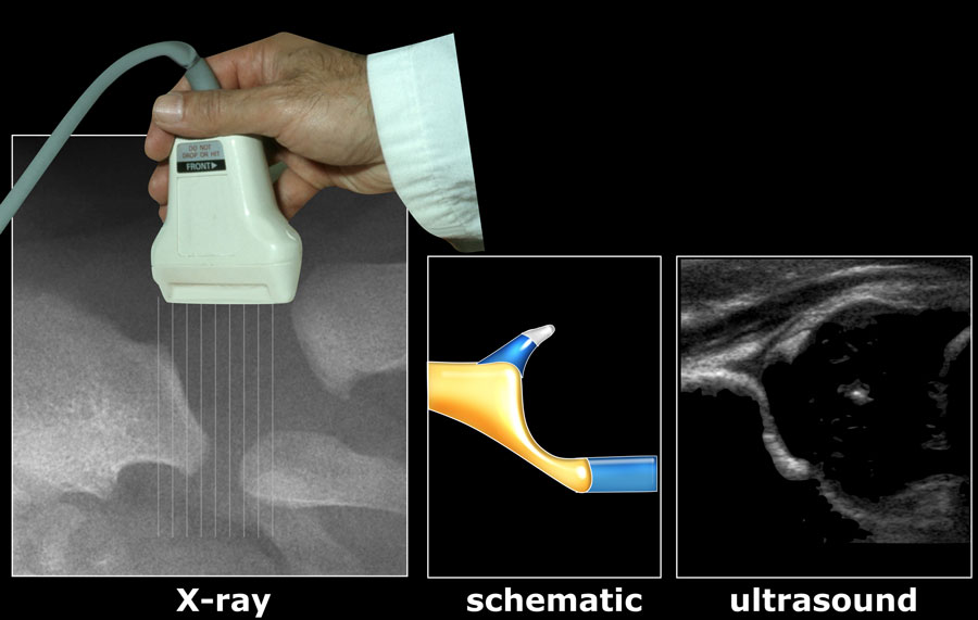 The Radiology Assistant Developmental Dysplasia Of The Hip
 Ultrasound Anatomy Of Sciatic Nerve In Popliteal Fossa Post
 Neonatal Cranial Ultrasound Standard Views
 Ultrasound Level One Services Heavenly 3d 4d Ultrasound
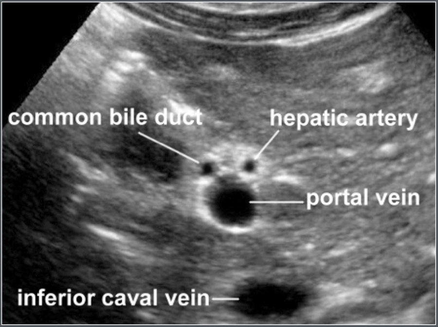
 Essential Ultrasound Anatomy
 Liver Measurement Ultrasound Pancreas And Its Proportions
 Shelby S 24 Week Ultrasound Anatomy Scan Mommymandy L
 Ultrasound Our Baby Girl K Y S L O T A N E T
 Just An Atlas Of Fetal Anatomy Ultrasound Handbook


Belum ada Komentar untuk "Anatomy Ultrasound"
Posting Komentar