Anatomy Of Foot Bones
The foot is located after the long shin bones and it starts from the back of your ankle to your toes. The bones of the feet are.
 Foot Anatomy Bones Ligaments Muscles Tendons Arches
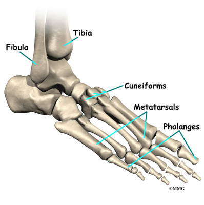
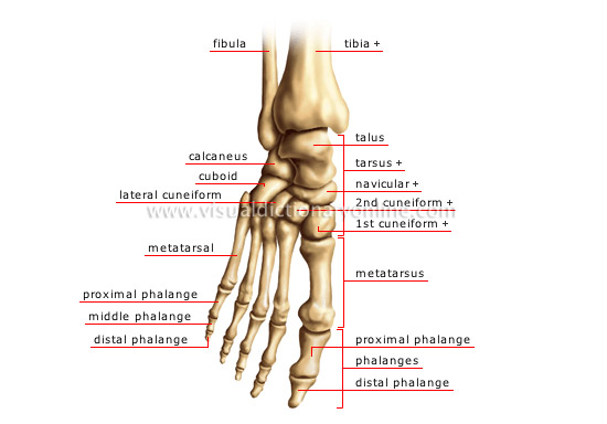
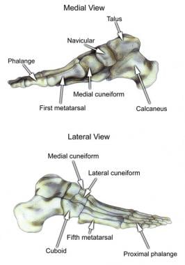
Talus the bone on top of the foot that forms a joint with the two bones of the lower leg.
Anatomy of foot bones. This unlabeled quiz of the bones of the foot will test your knowledge on how to label the structures of these bones. The calcaneus which is the bone in your heel. Learn this topic now at kenhub.
The different bones on each section of the foot. This enables them to evolve complex extraordinary hand and feet which they use for gripping grasping and rotating. The hindfoot consists of bone from the leg and the ankle joint.
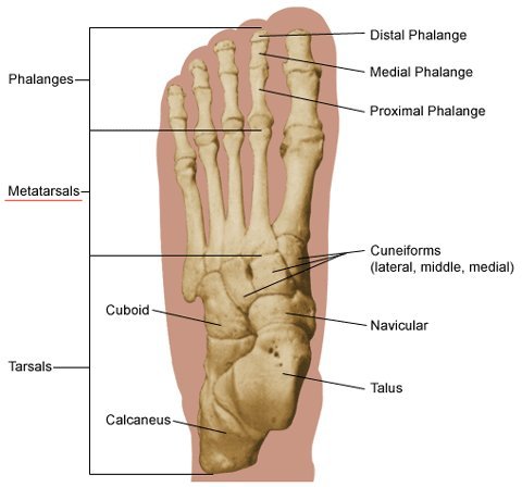
Anatomically the foot is divided into 3 sections. The hindfoot midfoot and the forefoot. The metatarsals which run through the flat part of your foot.
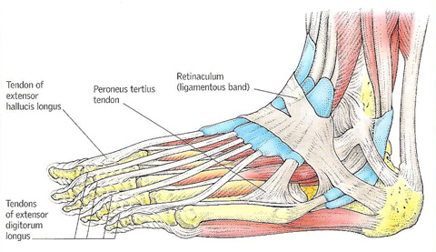
Hind means posterior so it basically the backward part of the foot. This is an article covering the articular surfaces ligaments and muscles that produce movement at the joints of the feet. You will be required to label the cuboid navicular calcaneus lateral cuneiform medial cuneiform medial cuneiform talus metatarsals and distalmiddleproximal phalanges.
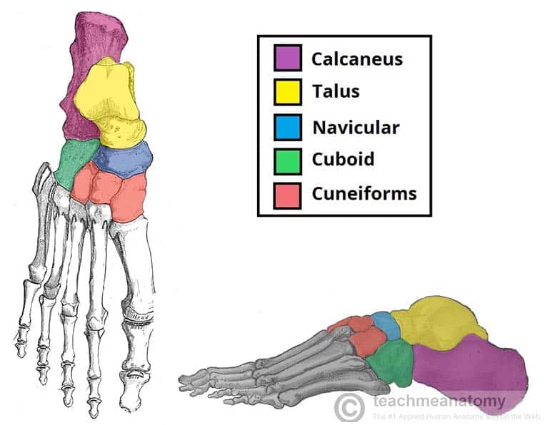
Foot anatomy bones the complex structure of human feet originates from grasping feet cells like and hand like that can be seen in primates today. The tarsal bones are 7 in number. The parts of the foot bones.
Foot bone quiz for anatomy and physiology. The phalanges which are the bones in your toes. The cuneiform bones the navicularis and the cuboid all of which function to give your foot.
This is an article covering the muscle attachments blood supply innervation and ossification of the phalanges of the foot. Calcaneus the largest bone of the foot which lies beneath the talus to form the heel bone. They are named the calcaneus talus cuboid navicular and the medial middle and lateral cuneiforms.
The talus bone supports the leg bones tibia and fibula forming the ankle. Tarsals five irregularly shaped bones of the midfoot that form the foots arch. The calcaneus is the largest of the tarsal bones located in the heel of the foot and bears the weight of the body as the heel hits the ground.
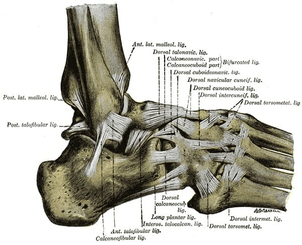
Bones and main ligaments of the foot. The talus which is the. The calcaneus heel bone is the largest bone in the foot.
The hindfoot is the posterior part of the foot. Our ancestors were tree dwellers and used to hang with all four limbs on branches. Tarsal bones gross anatomy.
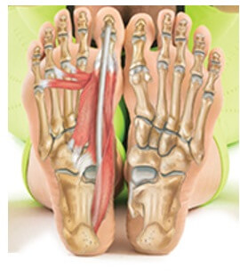
Parts of foot bones. Muscles tendons and ligaments run along the surfaces of.
 Bones Of The Foot Tarsals Metatarsals Phalanges
 Bones Of Foot Human Anatomy The Diagram Shows The Placement
 Anatomy Of Foot Bone Stock Illustration Illustration Of
 Foot Anatomy
 Bones The Of Foot
 Foot Ankle Anatomy Pictures Function Treatment Sprain Pain
 Bones Of The Foot Anatomy Tutorial
 Ball Of Foot Pain Do The Bottoms Of Your Feet Toes Hurt
 Foot And Ankle Anatomy Bones Muscles Ligaments Tendons
 Bones Of The Human Foot With The Name And Description Of All
 Ankle Foot Atlas Of Anatomy
Foot Anatomy
 Bones Of The Foot Illustrations Foot Anatomy Illustrations
 Cny Orthopedic Sports Medicine Pc Bodyzone Foot Ankle
 Anatomy Leg And Foot Bones

 Foot Bone Anatomy Overview Tarsal Bones Gross Anatomy
 Ankle Foot Atlas Of Anatomy
 Foot Ankle Anatomy Dr Sanford Bone And Joint Specialists
 Foot Vertebrate Anatomy Britannica
![]() Ankle And Foot Anatomy Bones Joints Muscles Kenhub
Ankle And Foot Anatomy Bones Joints Muscles Kenhub
 Human Anatomy Foot Bone
Belum ada Komentar untuk "Anatomy Of Foot Bones"
Posting Komentar