Cow Heart Anatomy
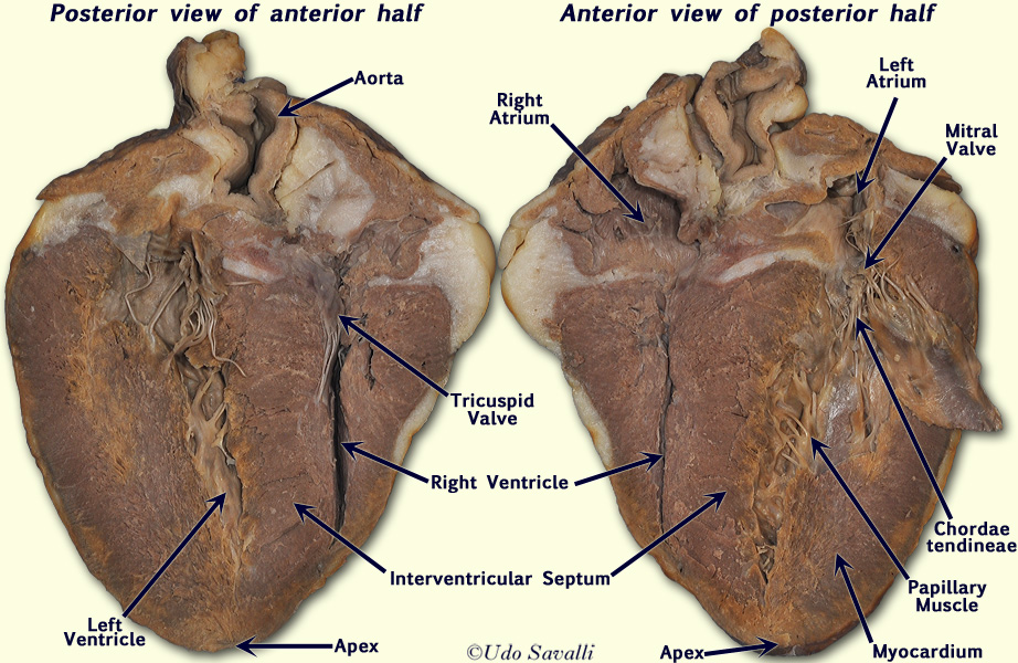
Chambers of the heart. They attach to the cusps of the atrioventricular valves also known as the mitral and tricuspid valves via the chordae tendineae and contract to prevent inversion or prolapse of these valves on systole or ventricular contraction.

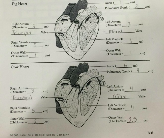
This difference in workload affects the anatomy of the ventricular walls.

Cow heart anatomy. Dissection of a fresh cow heart. Scientist cindy 46447 views. Both the cow and human heart serve the same purpose and the function of each heart is virtually identical.
The valves were probably cut when the heart was opened but if you follow the cords they should lead you to a thin flap that is the atrioventricular bicuspid valve. Anatomy of the cow heart. The muscular wall of the left ventricle being significantly thicker than that of the right.
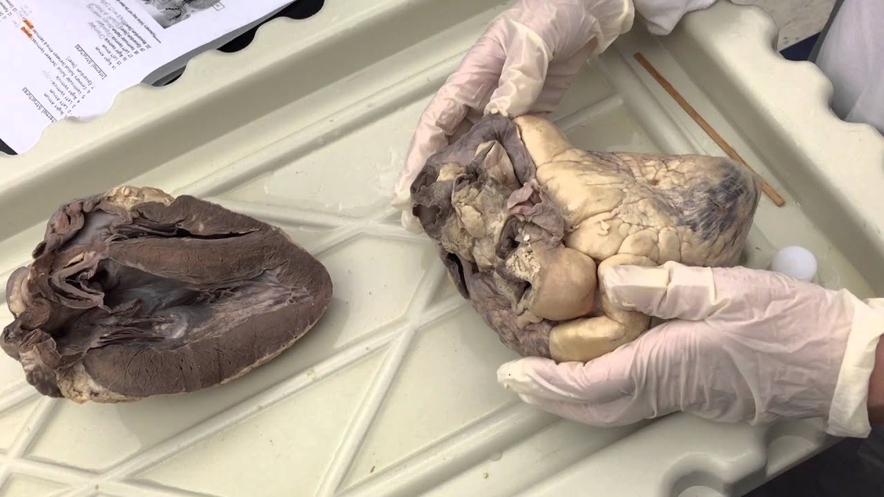
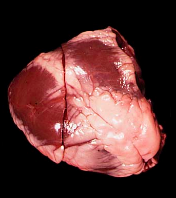
A cows mouth is adapted for grazing the top part of the mouth is a hard pad and the bottom part a row of flat topped teeth. These tendons hold the heart valves in place sometimes they are called the heartstrings. This photo shows the adult cow heart cut in cross section through the ventricles with the smaller right ventricle at top and the larger left ventricle at the bottom.
Left 9 papillary muscle does not show up too well. Below is a diagram of the anatomy of a cow. A human heart weighs between seven and 15 ounces whereas a cow heart can weight up to five pounds.
Oxygenated blood is brought from the lungs to the heart to be pumped through the body. Includes some review questions that show parts of the pig heart as well. We will explore the cow heart because its larger size makes it easier to see the important and complex structures of the heart.
Only the ventricles are visible in this photo. Consequently the left ventricle works harder than the right ventricle to maintain the same blood flow rate. Anatomy of the sheep brain video for anatomy class practice for the practical exam duration.
Cows have 32 teeth in all 8 incisors on the bottom part and 6 molars on the top and bottom parts on each side. What side of the heart is thicker than the other side. The two are separated by the interventricular septum and this example shows that the left ventricles walls are much thicker and stronger.
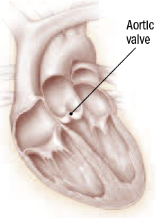
About pictured here is a cow heart with some incisions to show the chambers and valves inside. Note the amount of fatty material lightwhite colored tissue covering the surface of the heart. You can find a similar valve on the right side of the heart tricuspid.
 Cow Heart Structure Reading Industrial Wiring Diagrams
 Cow Heart Dissection
 Heart Dissection 8 Steps With Pictures
 Dissection Of Cow Heart In Anatomy And Physiology 2 Lab
 Bio202 Sheep Heart
 Nasco Cow Organ Beef Heart Dissection Science Education Materials Ls03717
 Anatomy And Physiology The Heart Dissection
 Sheep Heart Dissection Kit

 Heart Anatomy Heart Dissections Heart Labeling
 Sheep Heart Dissection Biology 2651 With Uyeno At Valdosta
 Cow Heart Anatomy Youtube
 Cow Heart Dissection Identification
 Lung Diagram Cow Reading Industrial Wiring Diagrams
 2nd Hour Anatomy Anatomy Of The Heart Sheep Pig And Cow
 Valve Replacement Mechanical Or Tissue Harvard Health

 Heart Anatomy Virtual Dissection

 Lung Diagram Cow Basic Electrical Wiring Theory

 Cow Heart Dissection Worksheet Sheep Heart Dissection
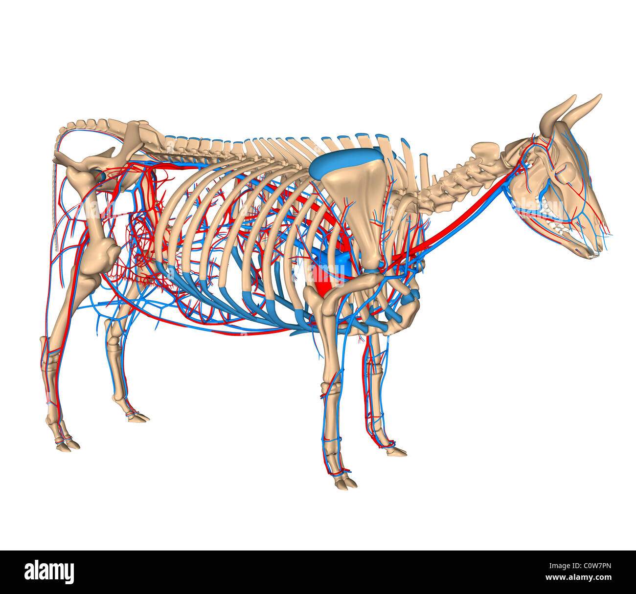 Anatomy Of The Cow Circulation Circulary Cor Heart Stock
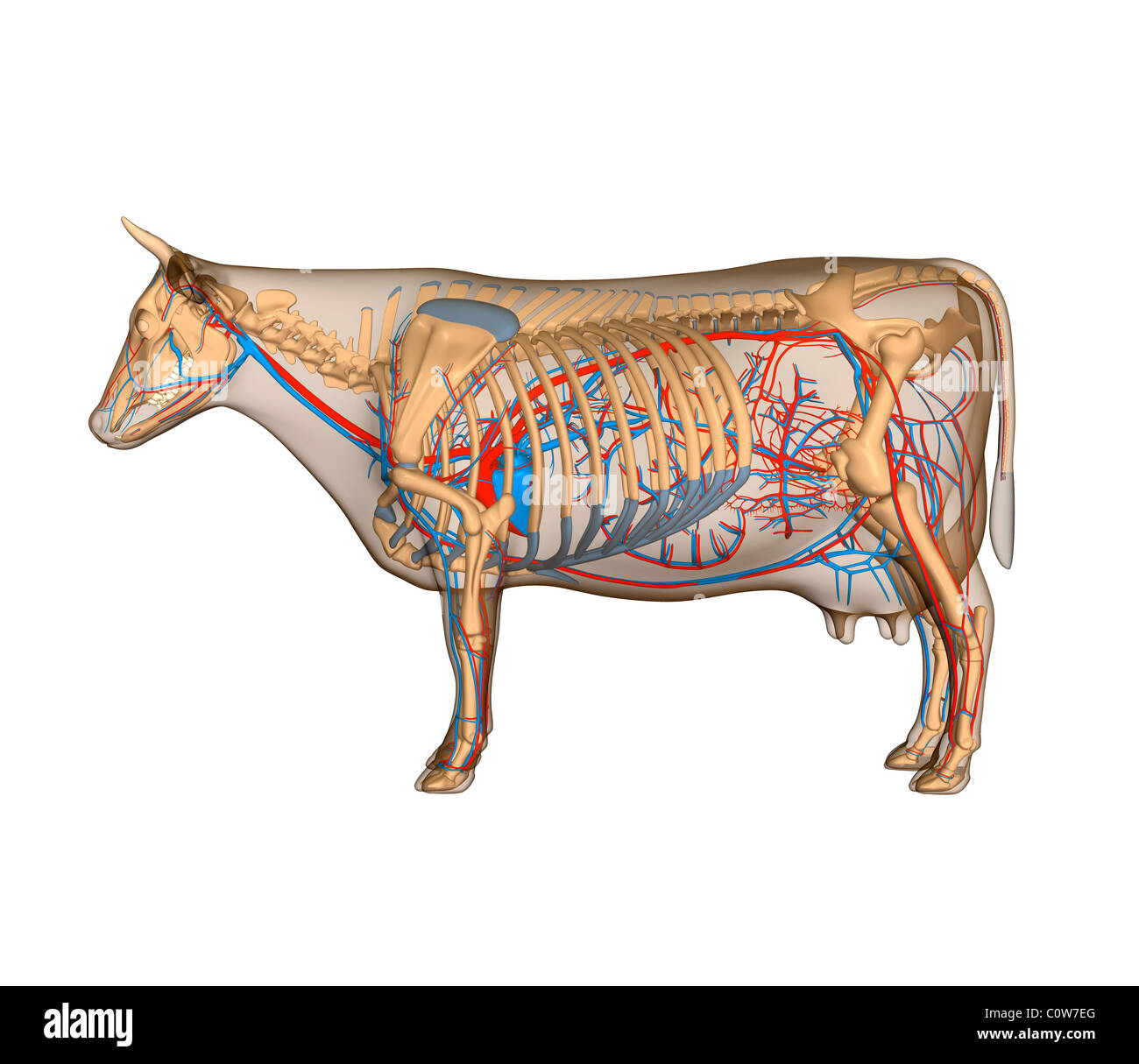 Anatomy Of The Cow Circulary Heart Circulation Stock Photo
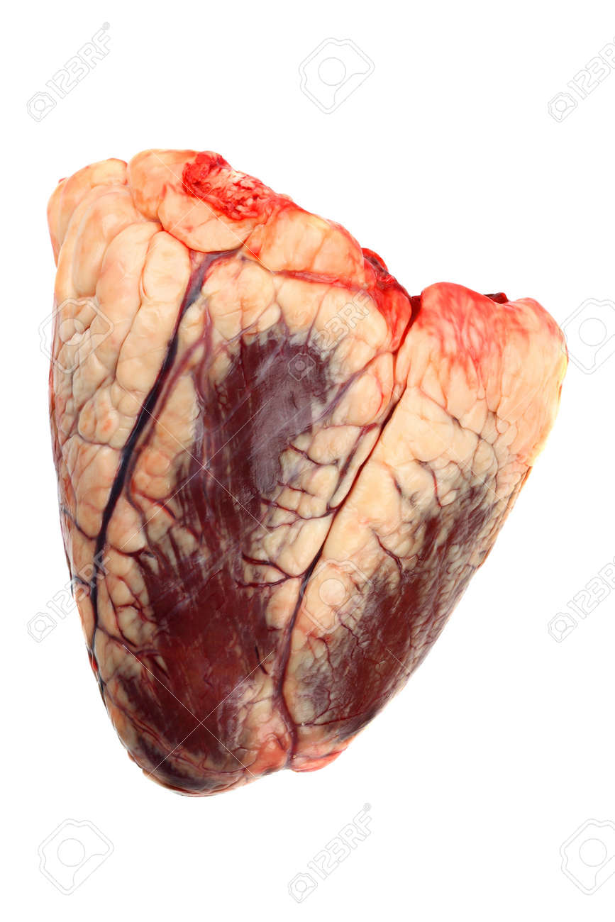 Raw Beef Heart Isolated On White
 Heart Anatomy Heart Dissections Heart Labeling
 Cow Heart Dissection Medicine Flashcards Quizlet
 Heart Ventricle Pin Labeled Anatomy Corner Anatomy
Belum ada Komentar untuk "Cow Heart Anatomy"
Posting Komentar