Chest Anatomy
It provides protection to vital organs eg heart and major vessels lungs liver and provides stability for movement. The epidermis is the outermost layer that provides a protective waterproof seal over the body.
 Chest Muscle Injuries Strains And Tears Of The Pectoralis
The thorax or chest is a part of the anatomy of humans and various other animals located between the neck and the abdomen.

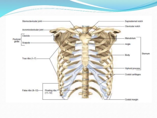
Chest anatomy. The shape of the chest is often regarded as potential insight into a disease process as in the case of barrel chest and respiratory dysfunction. The thorax includes the thoracic cavity and the thoracic wall. The chest wall is comprised of skin fat muscles and the thoracic skeleton.
It contains organs including the heart lungs and thymus gland as well as muscles and various other internal structures. Radiology basics of chest ct anatomy with annotated coronal images and scrollable axial images to help medical students and junior doctors learning anatomy. This page provides an overview of the chest muscle group.
Other anatomical structures such as the pleura only become clearly visible when abnormal. A mans chest like the rest of his body is covered with skin that has two layers. Learn about each of these muscles their locations functional anatomy and exercises for them.
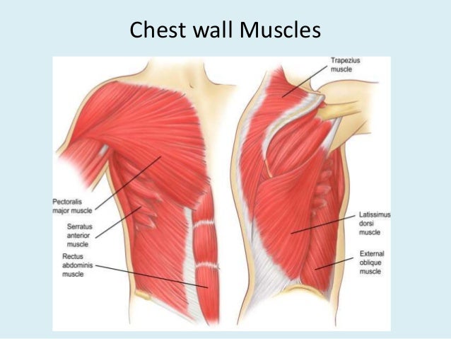
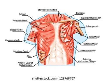
In fact some important structures such as the phrenic nerve are not visible at all. The anterior side of the thorax from the neck to the abdomen. The chest anatomy includes the pectoralis major pectoralis minor and the serratus anterior.
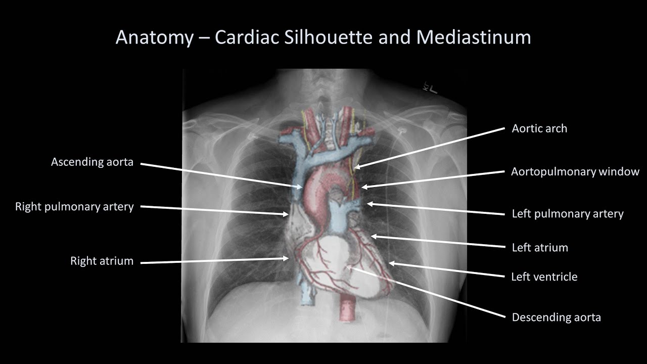
Chest x ray anatomy many structures of the chest are readily visible on a chest x ray but others are difficult to see.
 How To Interpret A Chest X Ray Lesson 2 A Systematic Method And Anatomy
 Surgical Anatomy Of The Chest Wall Thoracic Key


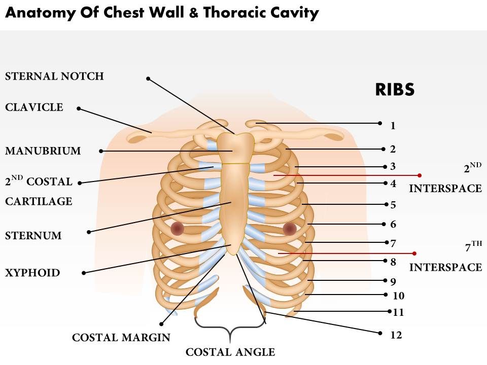 0514 Anatomy Of Chest Wall And Thoracic Cavity Medical
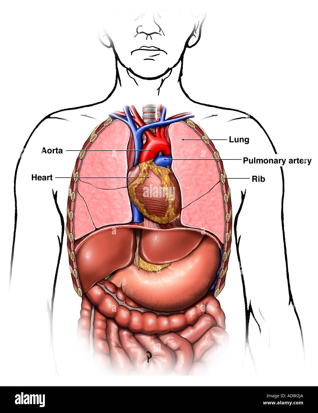 Anatomy Of The Thoracic Chest Organs Stock Photo 7711401
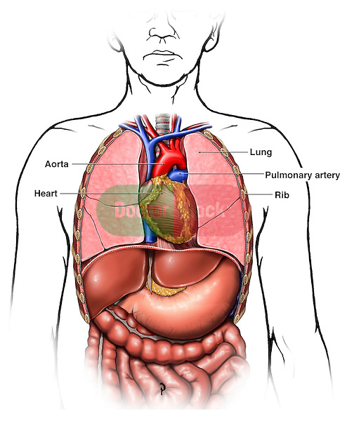
 Chest Anatomy Heart And Lungs
 Muscles Of The Chest And Upper Back
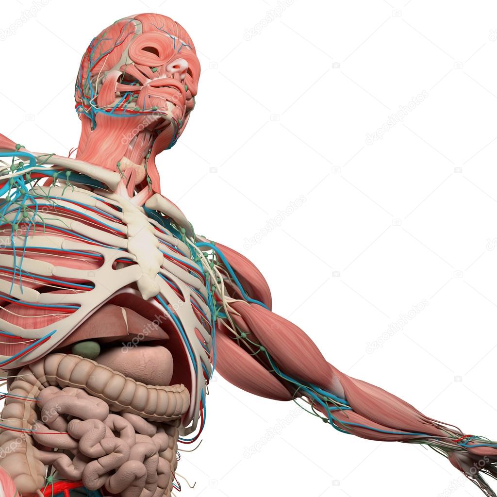 Human Anatomy Chest From Low Angle Bone Structure Veins
 Anatomy Of Thorax 2
 Anatomy Of The Chest Cavity Medical Art Works
 Human Chest Anatomy Images Stock Photos Vectors
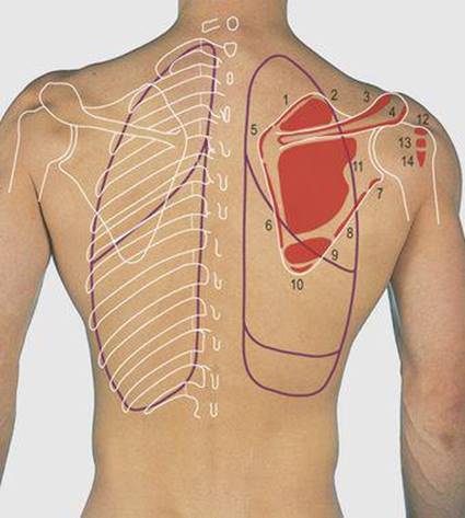 Thorax Surface Anatomy 4 Edition
![]() Chest Anatomy
Chest Anatomy
 Human Anatomy Female Human Anatomy Female Female Chest
 Radiological Anatomy Of Chest Including Lungs Mediastinum
 Opening The Thoracic Cavity
 Nerves Of The Chest And Upper Back
![]() Thorax Anatomy Wall Cavity Organs Neurovasculature
Thorax Anatomy Wall Cavity Organs Neurovasculature
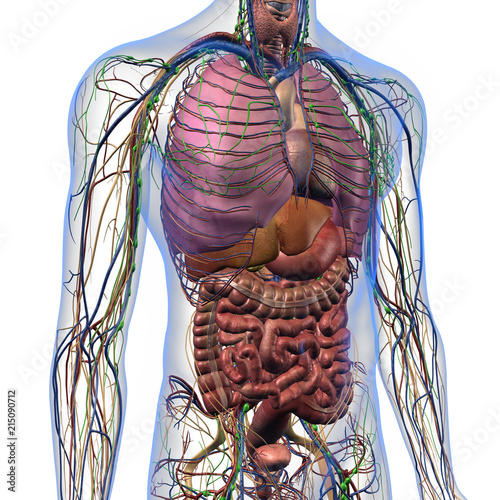 Male Internal Anatomy Of Chest And Abdominal Area On White
 Cardiomediastinal Anatomy On Chest Radiography Annotated
 Chest Bone Ribs Lung Heart Xiphoid Process Sternum Anatomy
 Anatomy Of The Thoracic Chest Organs Doctor Stock
 Thorax Chest Anatomy Archives Page 2 Of 6 Anatomy Note
![]() Normal Chest X Ray Anatomy Tutorial Kenhub
Normal Chest X Ray Anatomy Tutorial Kenhub
Stock Illustration
 Chest Anatomy Illustration Images Stock Photos Vectors
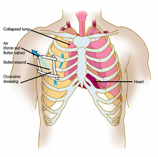 Anatomy Atlases Anatomy Of First Aid A Case Study Approach
 Thymus And Chest Anatomy In A Child 1866 Illustration
Belum ada Komentar untuk "Chest Anatomy"
Posting Komentar