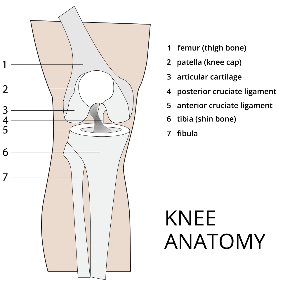
Knee Anatomy Acl
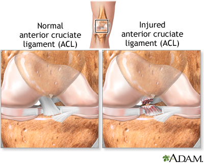
When the knee is fully extended both cruciate ligaments are taut and the knee is locked. The acl is responsible for a large part of the knees stability.
 Is An Acl Tear Worse Than A Meniscal Tear
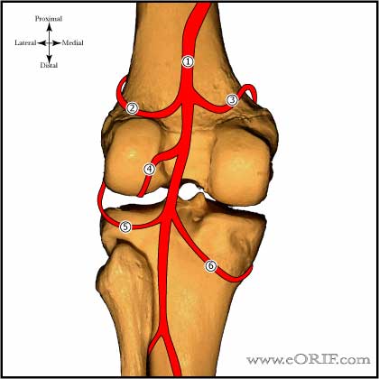
Gross anatomy the acl arises from the anteromedial aspect of the intercondylar area on the tibial plateau and passes upwards and backwards to.

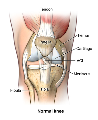
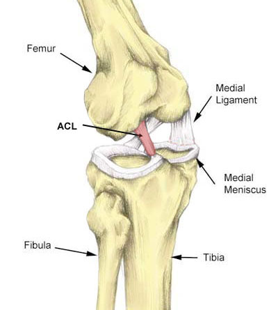
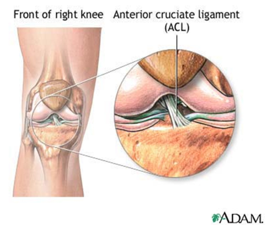
Knee anatomy acl. The cruciate ligament in the front of the knee is called anterior cruciate ligament or acl and the cruciate ligament in the back of the knee is called posterior cruciate ligament or pcl. The acl is one of the primary ligaments providing stability to the knee and therefore is one of the most commonly injured knee ligaments. The anterior cruciate ligament acl is a band of dense connective tissue which courses from the femur to the tibia.
Anterior cruciate ligament acl is one of the two cruciate ligaments that stabilize the knee joint. During movement of the knee the anterior cruciate ligament acl prevents anterior sliding of the tibia. The anterior cruciate ligament acl is one of a pair of cruciate ligaments the other being the posterior cruciate ligament in the human knee.
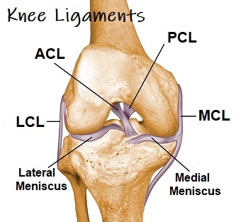
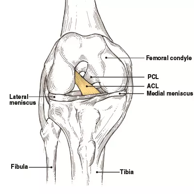
Pcl tears can cause pain swelling and knee instability. The 2 ligaments are also called cruciform ligaments as they are arranged in a crossed formation. The most common ligament injuries are acl tears mcl tears.
The acl is a key structure in the knee joint as it resists anterior tibial translation and rotational loads. The posterior cruciate ligament prevents posterior sliding of the tibia. Pcl posterior cruciate ligament strain or tear.
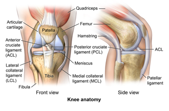
Anterior cruciate ligament acl resists anterolateral displacement of the tibia on the femur resists varus displacement at 0 degrees of flexion posterior cruciate ligament pcl. Ligaments are tough fibrous connective tissues which link bone to bone made of collagen. Cruciate ligaments this group of ligaments present inside the knee joint control the back and forth motion of the knee.
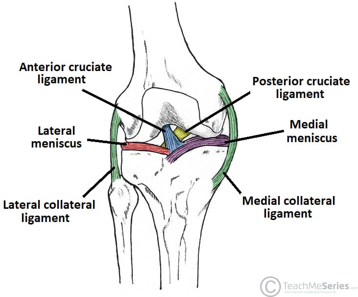
There is both an anterior cruciate ligament acl and a posterior cruciate ligament pcl. Acl anterior cruciate ligament strain or tear. The anterior cruciate ligament acl is one of a pair of ligaments in the center of the knee joint that form a cross and this is where the name cruciate comes from.
An acl tear often leads to the knee giving out and may require surgical repair. The acl and pcl are called cruciate ligaments because they form a cross in the middle of the knee joint. In knee joint anatomy they are the main stabilising structures of the knee acl pcl mcl and lcl preventing excessive movements and instability.
 Acl Solutions Acl Knee Anatomy And Diagram Images

 Knee Anatomy

 Deep And Superficial Mcl And Acl Double Bundle Anatomy
 15095 01a Right Knee Anatomy Exhibits
 Acl Injury Knee Specialist Chicago Westchester Hinsdale Il
 Acl Solutions Acl Knee Anatomy And Diagram Images
 How To Evaluate An Acl Injury 6 Steps
 Acl Anatomy Eorif

 Anterior Cruciate Ligament Acl Injury Medlineplus Medical
 Acl Tear Anterior Cruciate Ligament Sprain Diagnosis
 Knee Ligaments Cruciates Collaterals Knee Pain Explained
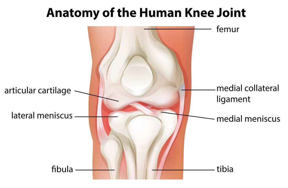
Anatomy Of The Knee Baxter Regional Medical Center
 All About Acl Injuries Girls Gone Strong
 Acl Solutions Acl Knee Anatomy And Diagram Images
 Can Stem Cells Treat An Acl Tear Or Torn Meniscus
 Acl Surgeons In Baton Rouge Acl Treatment Baton Rouge

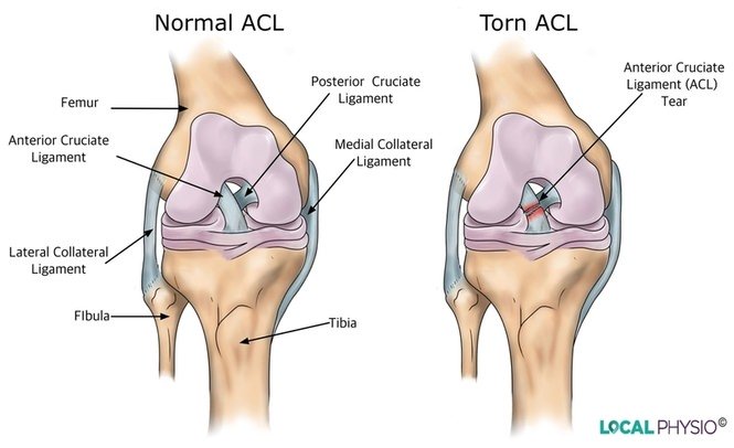 Anterior Cruciate Ligament Acl Injury Local Physio
Anterior Cruciate Ligament Acl Injuries Orthoinfo Aaos
 Acl Tear Brisbane Knee And Shoulder Clinic Dr Kelly
 Acl Tear Treatment Baltimore Md Towson Orthopaedic Associates

Common Knee Injuries Orthoinfo Aaos
 Acl Tear Q A Causes Diagnosing Treatment
 The Knee Joint Articulations Movements Injuries
 Biomechanics Functional Anatomy Behind Acl Tears Athletix
Belum ada Komentar untuk "Knee Anatomy Acl"
Posting Komentar