Hand Anatomy Palm
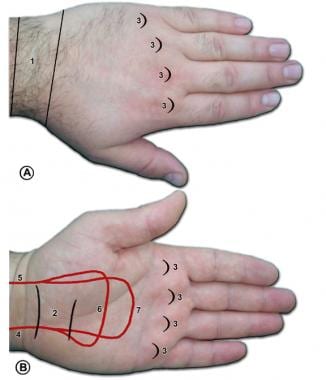
The major crease at the wrist on the palm side is simply called the wrist flexion crease. Superficialis tendons which pass through the palm side of the wrist and hand and attach at the bases of the middle phalanges.
Hand Anatomy Midwest Bone Joint Institute Elgin Illinois
The opisthenar area dorsal is the corresponding area on the posterior part of the hand.

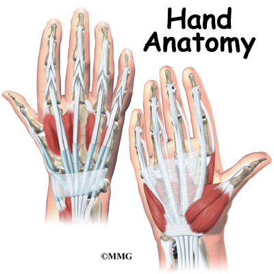
Hand anatomy palm. Also known as the broad palm or metacarpus it consists of the area between the five phalanges finger bones and the carpus wrist joint. All five metacarpals together are recognized as the metacarpus. Digits that extend from the palm of the hand the fingers make it possible.
The carpal bones are arranged in 2 interrelated rows. Other muscles in the palm attachments. Wrinkles the skin of the hypothenar eminence and deepens the curvature of the hand improving grip.
The palm side hand surface anatomy is described including names of creases in the fingers and palm. The heel of the hand is the area anteriorly to the bases of the metacarpal bones. One row connects with the ends of the bones in the forearm radius and ulna.
Located in the palm are 17 of the 34 muscles that articulate the fingers and thumb and are connected to the hand skeleton through a series of tendons. Wrist palm and fingers are thus made up of several small joints. The important structures of the hand can be divided into several categories.
They act with the profundus tendons to flex the wrist and mcp and pip joints. The wrist is a complex mechanical system of 8 small bones known as the carpal bones. The bones of the hand palm are known as metacarpal bones.
Bones muscles tendons nerves. The main tendons of the hand are. The wrist links the hand to the arm.
The hand can be considered in four segments. Anatomy of the hand and wrist. The palm comprises the underside of the human hand.
The palm volar which is the central region of the anterior part of the hand. All small carpal bones join the bones lying next to them and thus make the anatomy of the human hand very complex. Its formed as the wrist joint bends into a flexed position.
Areas of the human hand include. Another critical pair of terms used when describing sides of the hand or even wrist and forearm is radial and ulnar. The back of the hand shows the dorsal venous network a web of veins.
The front or palm side of the hand is referred to as the palmar side. Originates from the palmar aponeurosis and flexor retinaculum attaches to the dermis of the skin on the medial margin of the hand. This is the bottom of the body of the hand.
The back of the hand is called the dorsal side.
 Clinical Anatomy Hand Wrist Palmar Aspect Flexors
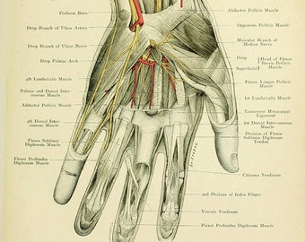 Human Anatomy Vintage Print Human Palm Anatomy Haeckel Human
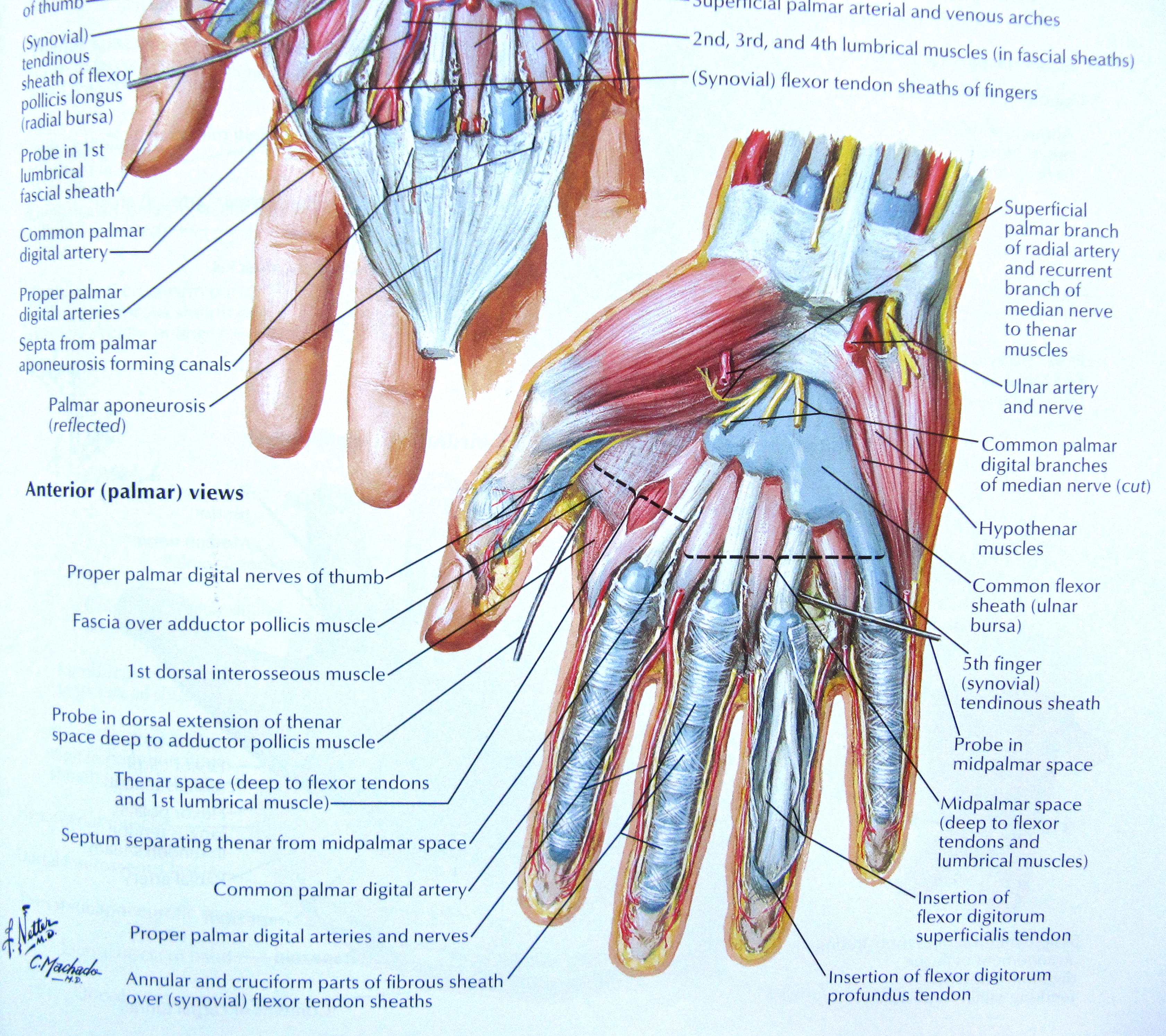 Notes On Anatomy And Physiology The Hand And The Tiger S
 Hand Pain Treatment Brooklyn Ny Wrist Pain Treatment Bronx
 10 Anatomy Of The Palm Of The Hand
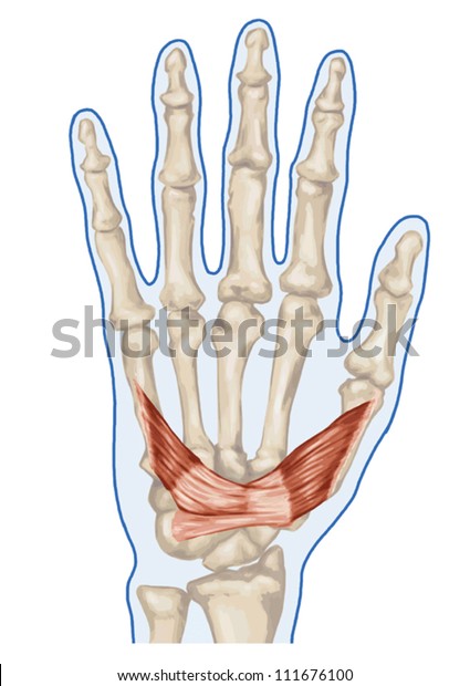 Anatomy Muscular System Hand Palm Muscle Stock Image
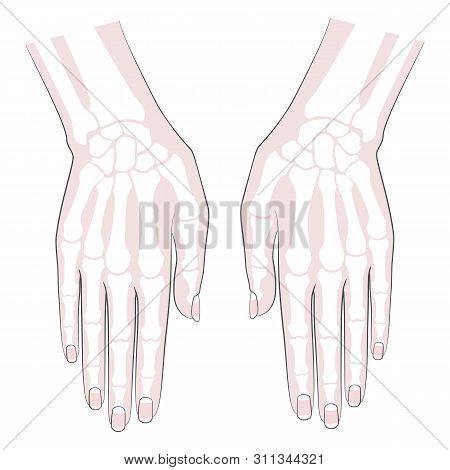 Anatomy Bones Hand Vector Photo Free Trial Bigstock
 Hand Anatomy Overview Bones Skin
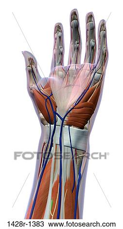 Female Palm And Wrist Anterior View Close Up Xray Skin
 95 Palm Of Hand Left Superficial Layer
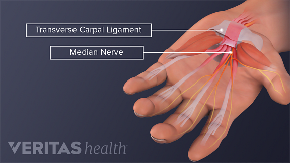 Is My Hand Pain From Carpal Tunnel Syndrome Or Something Else
 Muscles Of The Palm Hand For Anatomy Education Human Physiology
 Muscles Palm Hand Image Photo Free Trial Bigstock
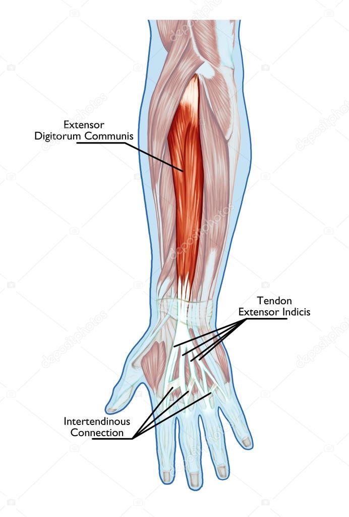 Anatomy Of Muscular System Hand Forearm Palm Muscle
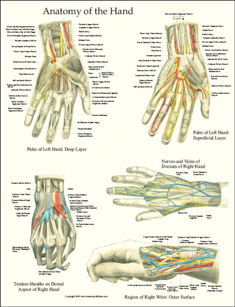 Hand And Wrist Anatomy Laminated Poster
 Muscles Of The Palm Hand For Anatomy Education Stock Photo
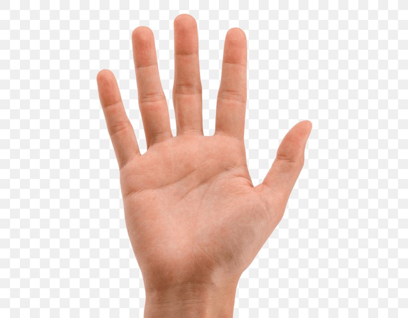 Finger Hand Palm Image Png 480x640px Finger Anatomy Arm
 Hand Surface Anatomy Palm Side
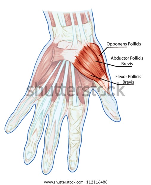 Anatomy Muscular System Hand Palm Muscle Stock Vector
David Nelson Hand Surgery Greenbrae Marin Hand Specialist
How Come When You Rest Your Hand Down Palm Facing Up Your
 Hand Muscles Palm Deep Labeled Muscular System Anatomy
 Hand Anatomy Level 6 This Is A View Of The Palm Side
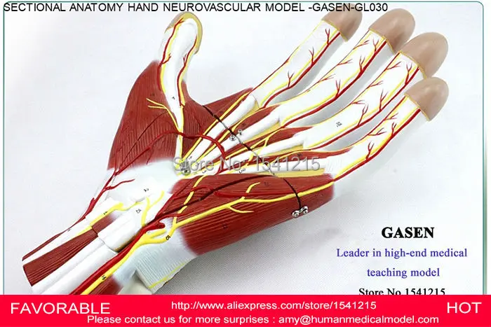 Us 260 0 Hand Sectional Anatomy Of Nerves And Blood Vessels Model Palm Anatomical Model Hand Anatomy Model Anatomical Model Gasen Gl030 In Medical



Belum ada Komentar untuk "Hand Anatomy Palm"
Posting Komentar