Tooth Anatomy Molar
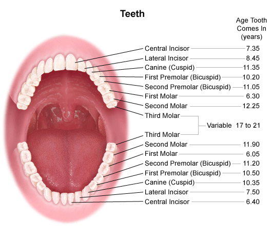
Canines 4 total. The root is the part of the tooth that extends into the bone and holds the tooth in place.
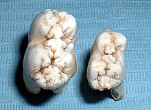
Management Of Third Molar Teeth From An Endodontic
The tissue composition of a tooth is only found within the oral cavity and is limited to the dental structures.

Tooth anatomy molar. The root canal is a passageway that contains pulp. Are also the largest teeth in the dentition. There are normally a total of 32 permanent secondary teeth in adults with 16 per jaw and eight in each quadrant which consists of distal to mesial 3.
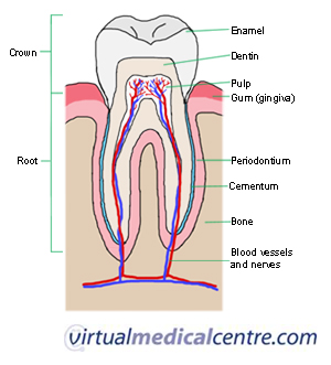
Enamel this is the outer and hardest part of the tooth that has the most mineralized tissue in. A normal adult mouth has 32 teeth which except for wisdom teeth have erupted by about age 13. Dentin this is the layer of the tooth under the enamel.
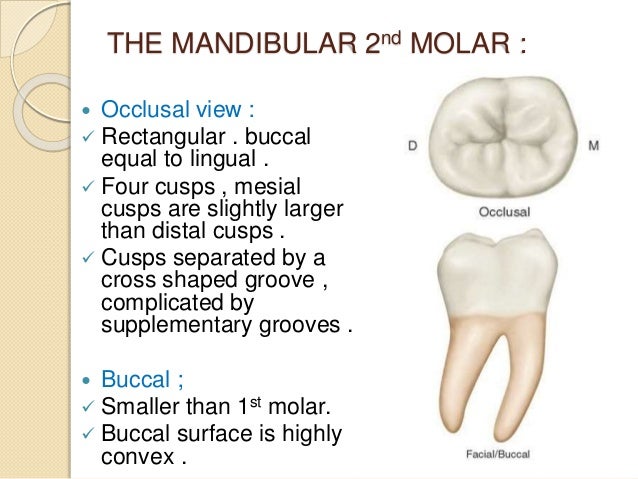
Premolars 8 total. The mandibular second molar is the tooth located distally from both the mandibular first molars of the mouth but mesially from both mandibular third molars. In humans the molar teeth have either four or five cusps.
10 maxillary 1st molar nbde part 1 boards study duration. Incisors 8 total. Each dental arch usually has six molars three in each quadrant if all have erupted.
The mandibular molars in particular the mandibular first molar are the most frequently endodontically treated teeth. This is true only in permanent teeth. These complexities include multiple canals isthmuses lateral canals and apical ramifications.
Permanent are the most posteriorly placed posterior teeth of the permanent dentition distal to the premolars. It makes up approximately two thirds of the tooth. Pass the dental boards 26577 views.
The middlemost four teeth on the upper and lower jaws. Here is an overview of each part. Their treatment offers a variety of anatomical challenges.
Each tooth is paired within the same jaw while the opposing jaw has teeth that are classified within the same category. Teeth between the canines and molars. Pulp this is the soft tissue found in the center of all.
In deciduous teeth the mandibular second molar is the last tooth in the mouth and does not have a third molar behind it. Anatomy of the tooth. Each tooth has several distinct parts.
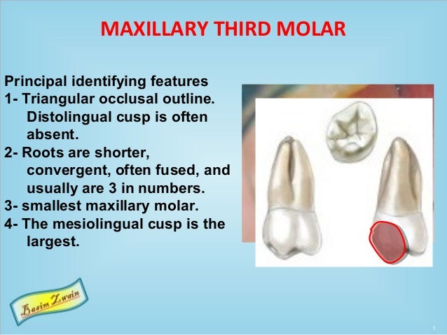
They are then progressively replaced by permanent secondary teeth from the age of six with the final eruption of the third molar between 18 24 years 5. The third rearmost molar in each group is called a wisdom tooth. However they are not grouped according to structure but rather by function.
Adult humans have 12 molars in four groups of three at the back of the mouth. The pointed teeth just outside the incisors. Its made up of several parts.
Also called cement this bone like material covers the tooths root.
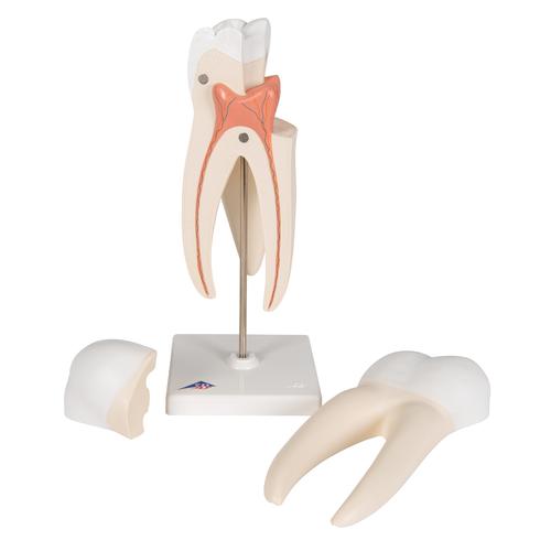 Upper Triple Root Molar Human Tooth Model 3 Part 3b Smart Anatomy
 Dental Anatomy Maxillary Molars
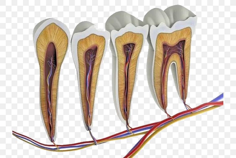 Human Tooth Anatomy Molar Pulp Png 733x550px Watercolor
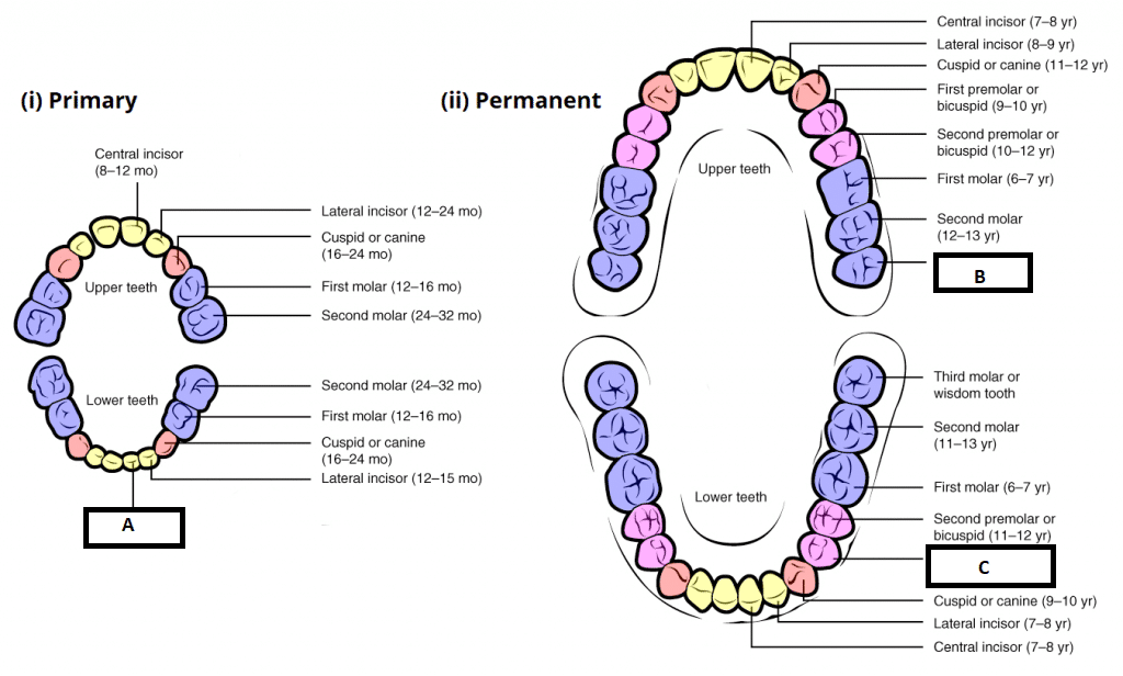 Child And Adult Dentition Teeth Structure Primary
Vc Dental Tooth Anatomy Education
 Molar Anatomy Shared By Dr Gregory Bowen San Antonio
 Amazon Com Dental Teeth Anatomy Structure Molar Incisor 2
Pediatric Center Penn State Hershey Medical Center Tooth
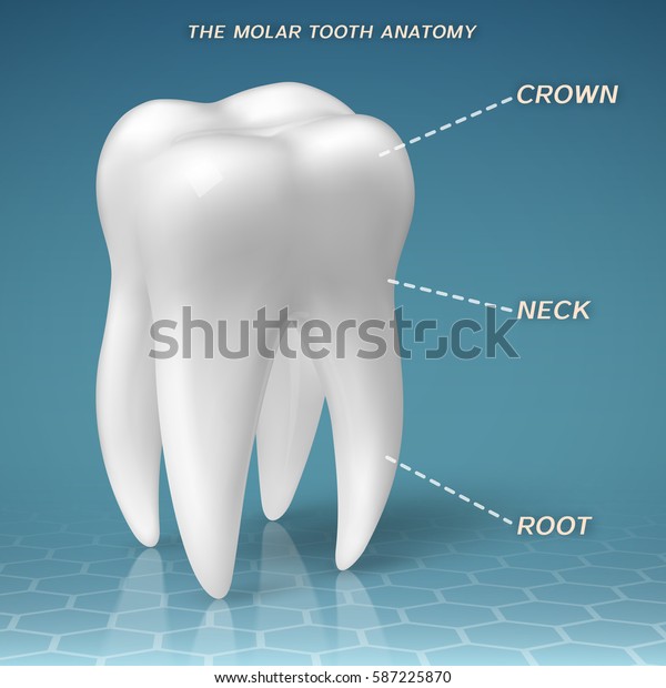 Molar Anatomy Crown Neck Root Tooth Stock Vector Royalty
 Tooth Anatomy Section Of A Human Molar
 Anatomy And Development Of The Mouth And Teeth
The Permanent Mandibular Molars Dental Anatomy Physiology
 Teeth Anatomy Adult Teeth Permanent Dentition
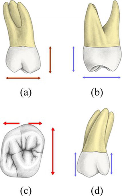 External And Internal Root Canal Anatomy Of The First And
The Permanent Mandibular Molars Dental Anatomy Physiology
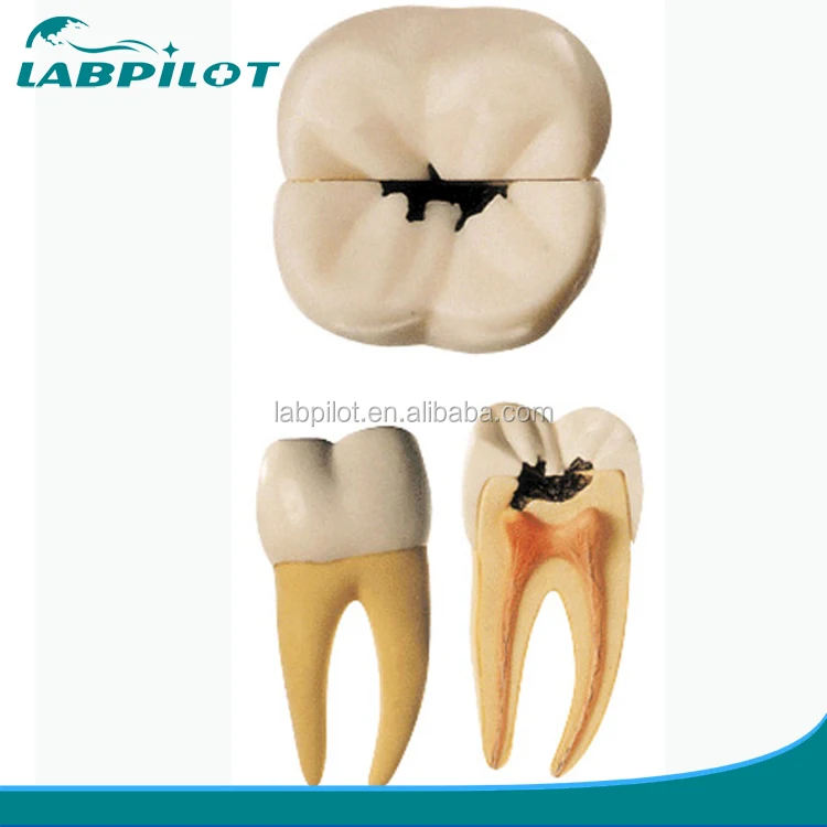 Molar Tooth Decay Anatomical Model Dental Caries Model Buy Dental Caries Tooth Decay Molar Product On Alibaba Com
Human Being Anatomy Teeth Cross Section Of A Molar
The Permanent Mandibular Molars Dental Anatomy Physiology
Human Being Anatomy Teeth Cross Section Of A Molar
 Tooth Anatomy Everything You Should Know About The Teeth
 Human Tooth Anatomy Vector Diagram Of Healthy Molar
Management Of Third Molar Teeth From An Endodontic



Belum ada Komentar untuk "Tooth Anatomy Molar"
Posting Komentar