Xray Elbow Anatomy
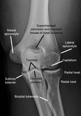
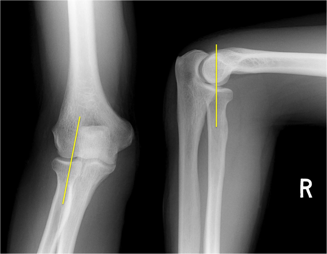
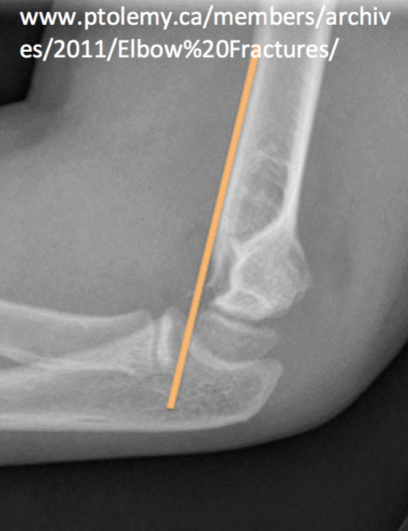
Drawn down the anterior surface of the humerus should intersect the middle 13 of the capitellu. The chronic valgus overload can cause an osteochondral lesion on the lateral side of the elbow.
 Ecr 2015 C 2327 Commonly Missed Fractures In The
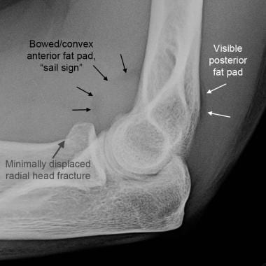
It is caused by displacement of the fat pad around the elbow joint.
Xray elbow anatomy. Elbow fat pads there are pads of fat close to the distal humerus anteriorly and posteriorly. Test your knowledge about elbow xray anatomy with this online quiz. Normal elbow x ray appearances on the lateral image there is often a visible triangle of low density lying anterior to the humerus.
Systematic review whenever you look at an adult elbow x ray review. Capitellum of the humerus with the ra. Alignment fat pads bone cortex alignment check the anterior humeral line.
It is the result of repetitive impaction and shear forces. There is a focal lucency in the capitellum and some fragmntation. Stanford bone tumor bayesian network issssr msk lectures for residents ocad msk cases from around the world stanford msk mri atlas has served almost 800000 pages to users in over 100 countries.
The radiograph is of a 15 year old baseball player with 4 year history of elbow pain and a recent episode of locking. Use the mouse to scroll or the arrows. Injuries around the joint can produce a joint effusion which will displace the fat pads making them more visible.
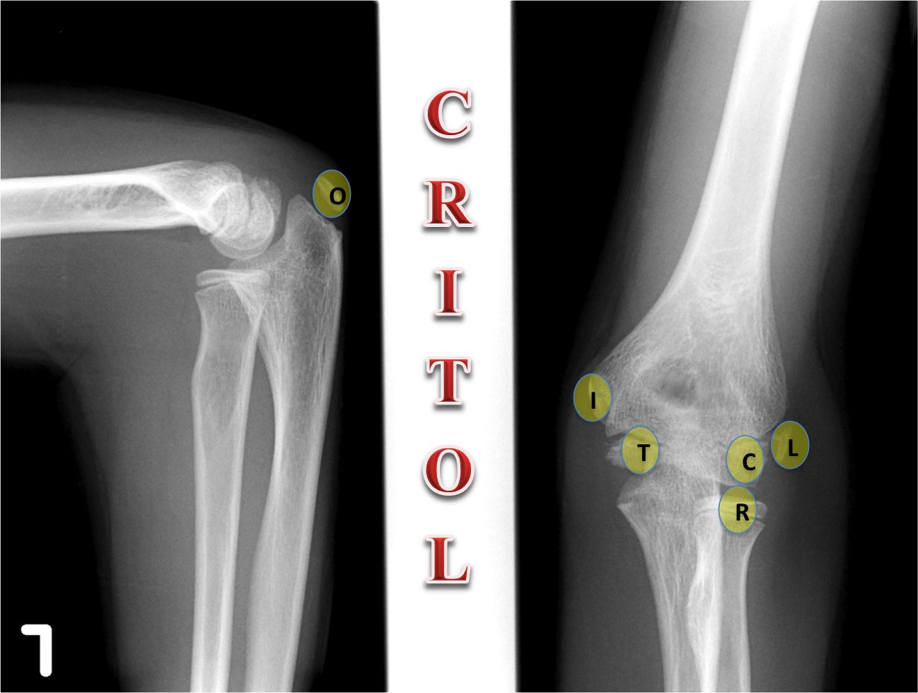
On an elbow x ray a fat pad sign suggests an occult fracture. A trivia quiz called elbow xray anatomy. They are extrasynovial but intracapsular.
Knee shoulder shoulder arthrogram ankle elbow wrist hip contact. On a normal elbow x ray only a small stripe of an anterior fat pad should be visible. This is a normal structure.
The anterior fat pad is seen in most but not all normal elbows. Both anterior and posterior fat pad signs exist and both can be found on the same x ray. Copyright c 2005 2019 alex freitas md.
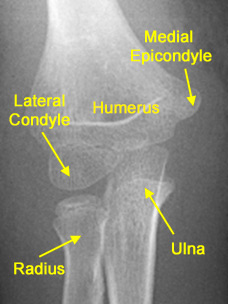
This is the anterior fat pad which lies within the elbow joint capsule. No posterior fat pad should be seen. The elbow is a complex synovial joint formed by the articulations of the humerus the radius and the ulna.
Gross anatomy articulations the elbow joint is made up of three articulations 23.
 Imaging Of Elbow Fractures And Dislocations In Adults
 Startradiology
 File X Ray Of Normal Elbow By Lateral Projection Jpg Wikipedia
 Normal Radiographic Anatomy Of The Elbow Radiology Case
 Imaging Of Elbow Fractures And Dislocations In Adults
 Film Xray Elbow Radiograph Show Normal Stock Photo Edit Now
 Interpreting Elbow And Forearm Radiographs Taming The Sru
 Orthokids Elbow Fractures
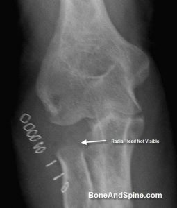 Xrays Of Elbow Injuries Bone And Spine
 Olecranon Bursitis Symptoms And Treatment The Hand Society
 Elbow Xray Interpretation
 Radiographic Anatomy Of The Skeleton Elbow
 Startradiology
 Startradiology
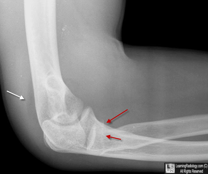 Learning Radiology Posterior Fat Pad Sign
 Startradiology
 Film Critique Of The Upper Extremity Part 2 Elbow And Forearm
The Elbow
 Xray Elbow Stock Photo Download Image Now Istock
 Image Iq 12 Year Old Boy With Acute Medial Elbow Pain
 Elbow Anatomy Pictures Bones Muscles Nerves
Elbow Radiographic Anatomy Wikiradiography
 The Elbow
 Ap Elbow Radiograph Anatomy Diagram Quizlet
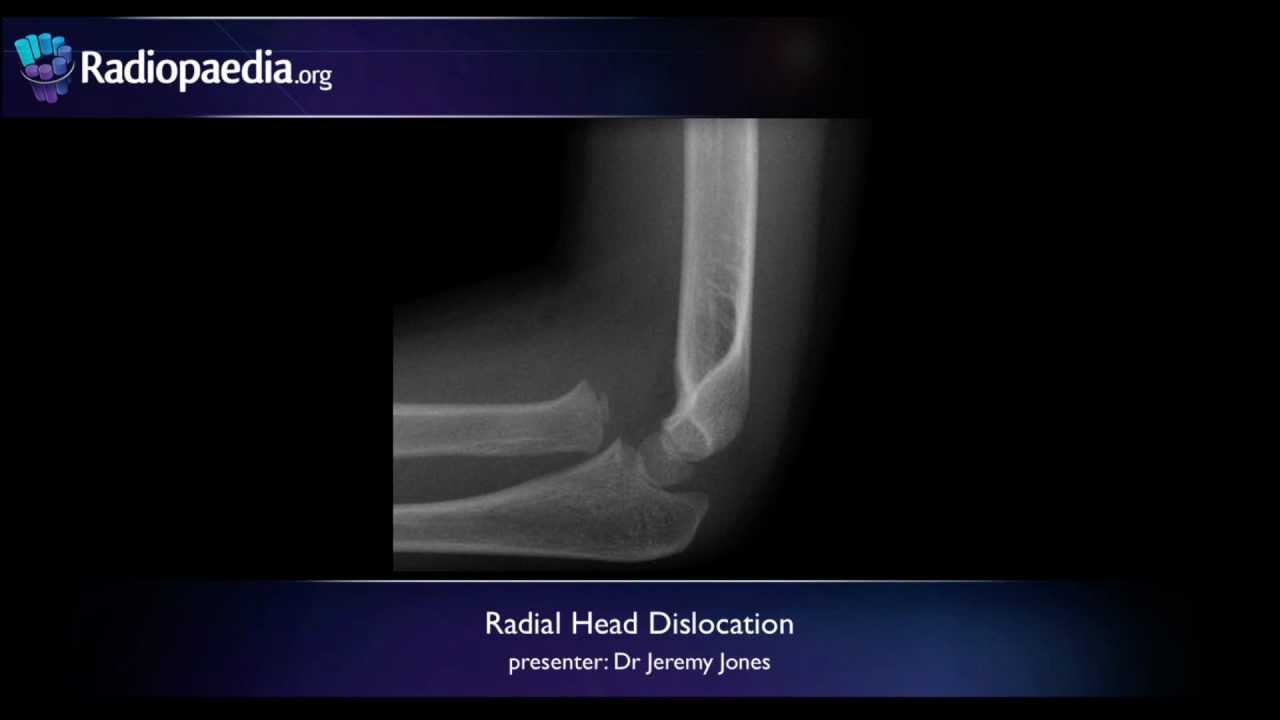 Radial Head Dislocation Radiology Video Tutorial X Ray
 Interpreting Elbow And Forearm Radiographs Taming The Sru


Belum ada Komentar untuk "Xray Elbow Anatomy"
Posting Komentar