Heart Valve Anatomy
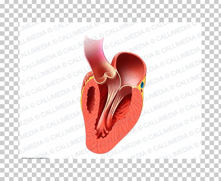
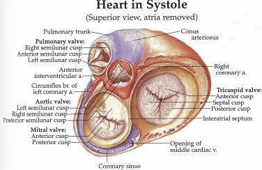
Semilunar valves control blood flow out of your heart. The external surfaces of the valves are covered by endocardium.
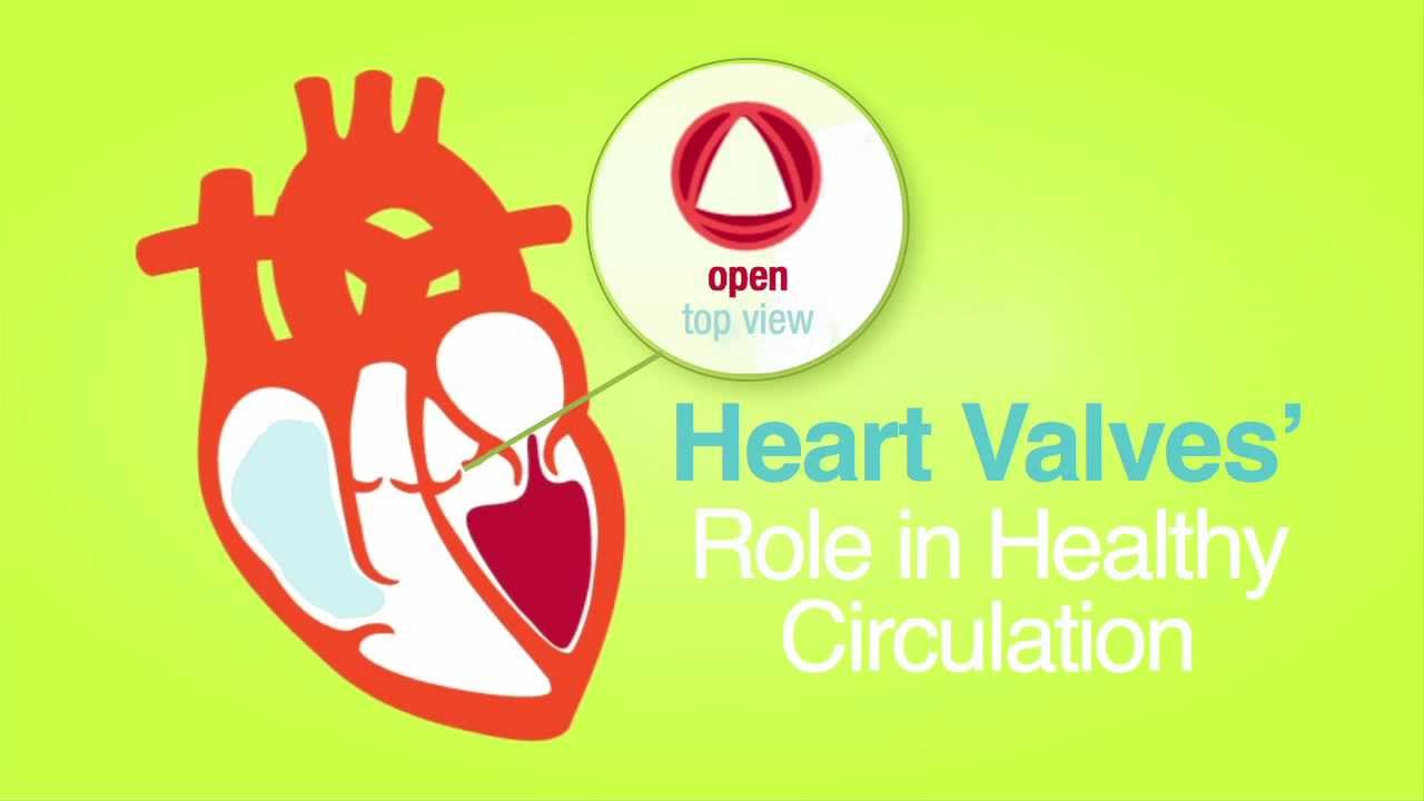
 Roles Of Your Four Heart Valves American Heart Association
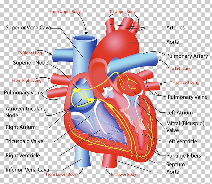
The right side of the heart pumps blood through the lungs where it picks up oxygen.
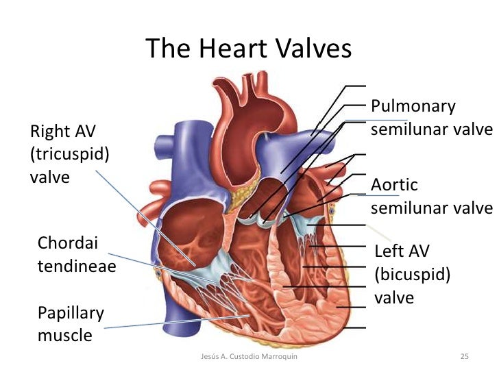
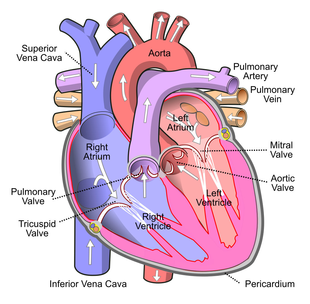
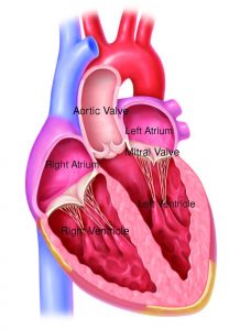
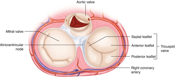
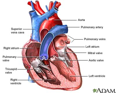
Heart valve anatomy. The valve between the right atrium and the right ventricle is called the tricuspid valve. The valve between the left atrium and the left ventricle is called the mitral valve. Atrioventricular av valves tricuspid valve.
They are composed of connective tissue and endocardium the inner layer of the heart. Valve in anatomy any of various membranous structures especially in the heart veins and lymph ducts that function to close temporarily a passage or orifice permitting movement of a fluid in one direction only. Your heart has four chambers and four valves that regulate blood flow.
The four valves of the heart. The heart is one of the most important organs in the body. Understanding heart valves anatomy is important in grasping the overall function of the heart.
The right atrioventricular valve or av valve controls blood flow from the right atrium to the right ventricle. This heart valve is located between the left atrium and left ventricle. A valve may consist of a sphincter muscle or two or three membranous flaps or folds.
This heart valve is located between the right atrium and the right ventricle. Introduction to the anatomy of the heart valves. When closed it allows the.
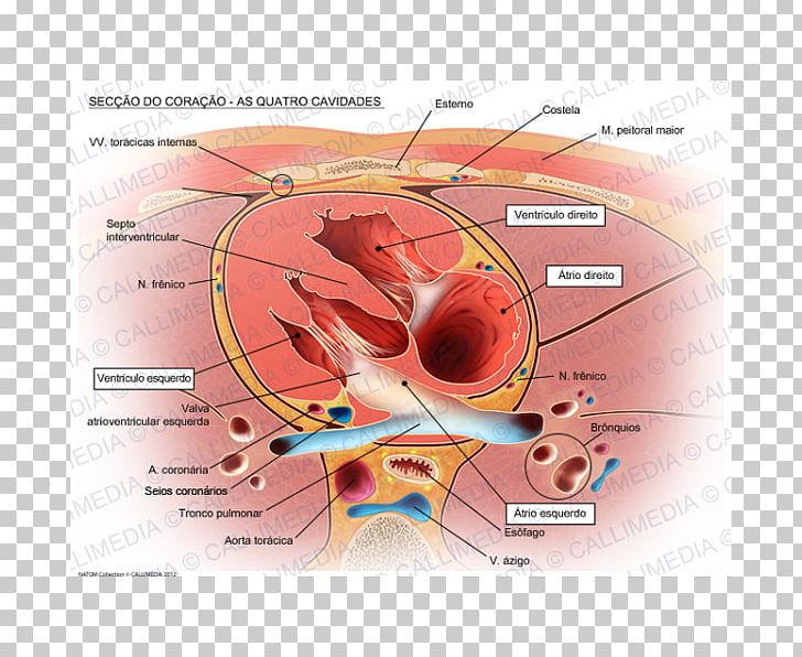
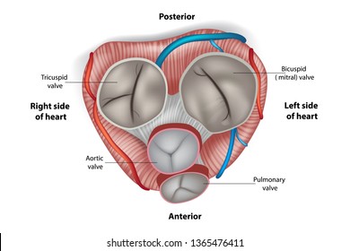
The two atrioventricular av valves the mitral valve bicuspid valve and the tricuspid valve which are between the upper chambers atria and the lower chambers ventricles. The valves of the heart are structures which ensure blood flows in only one direction. When closed it allows oxygen depleted blood returning to.
The left side of the heart receives the blood containing oxygen and pumps the blood to the rest of your body. The four valves in the mammalian heart are. The valves of the heart are structures which ensure blood flows in only one direction.
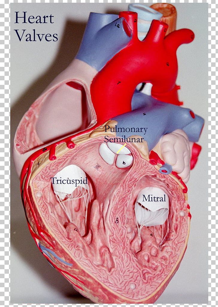
They are composed mostly of fibrous connective tissue that extends from the heart walls. The two semilunar sl valves the aortic valve and the pulmonary valve. Blood flows out of the right ventricle to the lungs through the pulmonary valve.
It is responsible for propelling blood to every organ system including itself.
 Heart Valves Anatomy Mitral Valve Pulmonary Valve Aortic Valve
 Heart Anatomy
:max_bytes(150000):strip_icc()/heart_inner_section-577d5c673df78cb62c939314.jpg) Atria Of The Heart Function
Atria Of The Heart Function
 Heart Valve Human Anatomy And Physiology 2 Circulatory
Heart Anatomy Anatomy And Physiology Openstax
 Heart Valve Right Atrium Aorta Anatomy Png Clipart Anatomy
![]() Heart Valves Anatomy Tricuspid Aortic Mitral Pulmonary Kenhub
Heart Valves Anatomy Tricuspid Aortic Mitral Pulmonary Kenhub
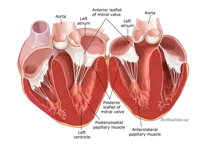
 Mitral Valve 1 Anatomy Online Learning
 Aortic Valve Images Stock Photos Vectors Shutterstock
 Anatomy Of The Heart Heart Valves Function Purpose And How
 The Heart Valves With Heart Circulatory Premium 2
 Anatomy Of The Human Heart
 Heart Anatomy Internal Medical Art Library
 Heart Valve Circulatory System Anatomy Human Body Png
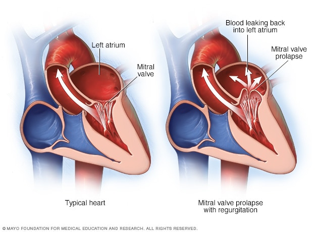
 Learn About Bicupsid Aortic Valves And Mitral Valve Prolapse
 Heart Valve Anatomy
 Transcatheter Mitral Valve Repair First Coast Heart
 Anatomy Of The Tricuspid Valve And Pathophysiology Of
Roles Of Your Four Heart Valves American Heart Association
 Heart Valve Anatomy Tricuspid Valve Png Clipart Anatomy
 Heart Model With Valves
 Heart Valves Anatomy
![]() Heart Valves Anatomy Tricuspid Aortic Mitral Pulmonary Kenhub
Heart Valves Anatomy Tricuspid Aortic Mitral Pulmonary Kenhub
 Heart Valves Function And Anatomy How The Heart Valves Work
 Heart Valve Stock Photos Heart Valve Stock Images Alamy
 Heart Valve Surgery Sciencedirect
 What Do I Need To Know About Heart Valves Lesson
 Mitral Valve Disease Symptoms And Causes Mayo Clinic

 Heart Valve Anatomy Purposegames
Belum ada Komentar untuk "Heart Valve Anatomy"
Posting Komentar