Renal Vein Anatomy
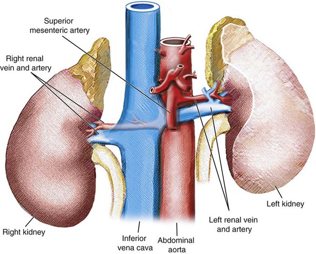
Renal vein s lie in front of the corresponding renal artery. There are many clinically significant anatomical variants of the course of each renal vein as well as anatomical variants of the venous tributaries that drain into the renal veins.
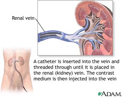 Renal Veins Medlineplus Medical Encyclopedia Image
The posterior veins assist in draining the back section of each kidney while the anterior veins assist the front part.
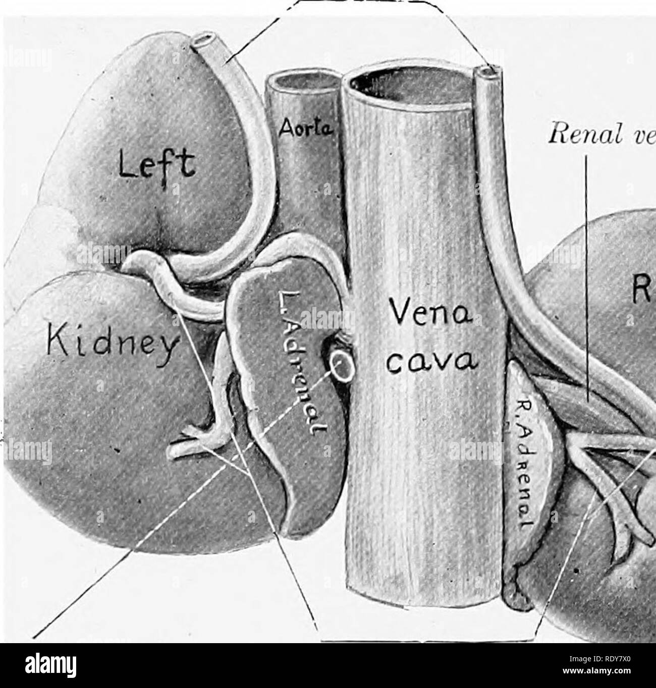
Renal vein anatomy. Gross anatomy course the renal vein is formed by the union of two to three renal parenchymal veins in the renal sinus. Each renal vein drains into a large vein called the inferior vena cava ivc which carries blood directly to the heart. The left renal vein is longer than the right due to the position of the ivc on the right side of the body and also drains venous blood from the.
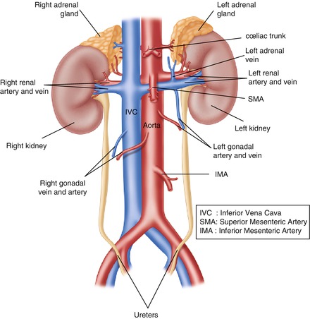
The right renal vein receives tributaries exclusively from the kidney while the left receives blood from a number of other organs as well. Each kidney is drained by its own renal vein the right and left renal vein. Some of these are specific to the left renal vein.
The renal veins are blood vessels that return blood to the heart from the kidney. There are two renal veins a left and a right. The renal veins are asymmetric paired veins that drain the kidneys.
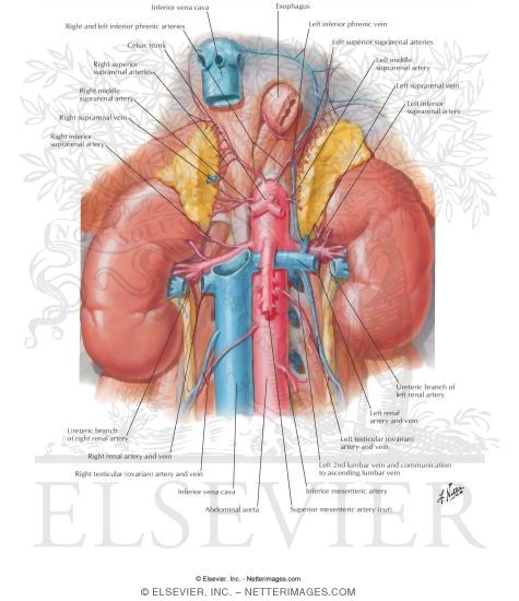
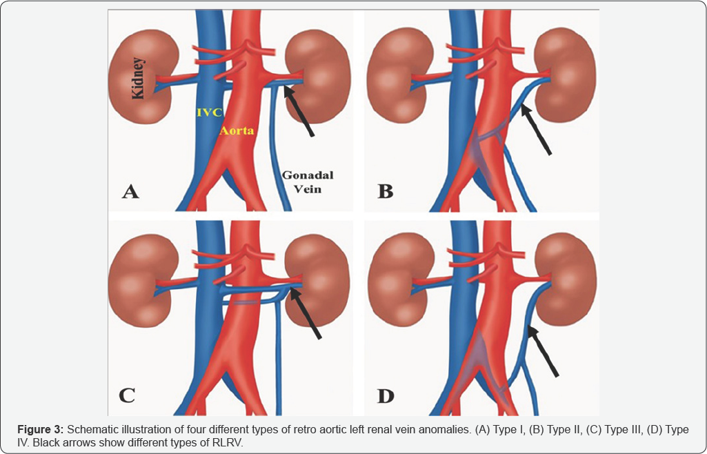
The right supra renal vein terminates directly in the inferior vena cava as. Left renal vein anomalies are generally classified into four types 2. The renal veins are veins that drain the kidney.
The renal veins form from the confluence of the interlobar veins and empty into the inferior vena cava ivc which empties back into the right side of the heart. The right suprarenal vein drains into the inferior vena cava and the left suprarenal vein drains into the left renal vein. They connect the kidney to the inferior vena cava.
As they enter the kidneys each vein separates into two parts. Venous drainage is provided by the suprarenal veins. They branch off the inferior vena cava and drain oxygen depleted blood from the kidneys.
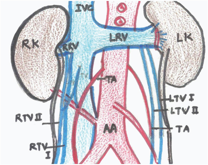
There are several variations in renal venous anatomy. B transverse section of the kidney showing relative position of vascular structures in the renal pelvis. Type i the ventral pre aortic limb of the left renal vein is obliterated but the dorsal retro aortic limb persists and joins the ivc in the normal position.
A there is extensive communication between the renal venous plexus and lumbar gonadal and adrenal veins which provide alternative outflow in the setting of renal vein thrombosis particularly on the left. The renal veins are not symmetrical as the left and right renal veins have significantly different courses as they travel toward the inferior vena cava draining the blood from each kidney as well as other organ systems such as the gonads adrenal glands and diaphragm. It emerges from the renal hilum anterior to the renal a.
Clinical anatomy for dummies. They carry the blood filtered by the kidney. Lymph is drained into the lumbar lymph nodes.
 Renal Vein Wikipedia
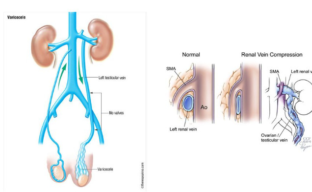 Testicular Vein Nutcracker Syndrome 네이버 블로그
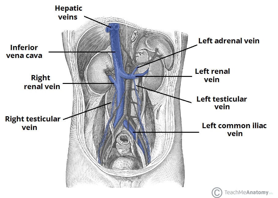 Venous Drainage Of The Abdomen Teachmeanatomy
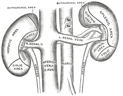
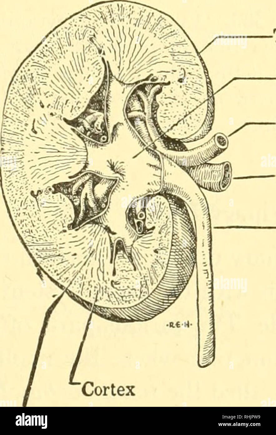 Biology Of The Vertebrates A Comparative Study Of Man And
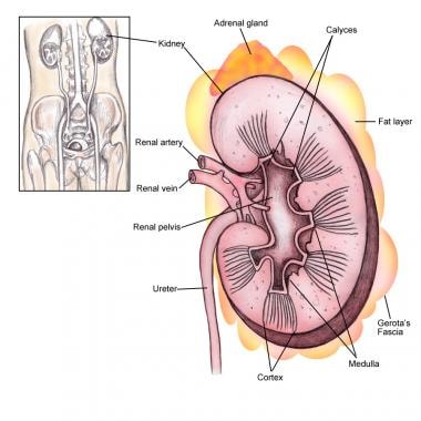 Kidney Anatomy Overview Gross Anatomy Microscopic Anatomy
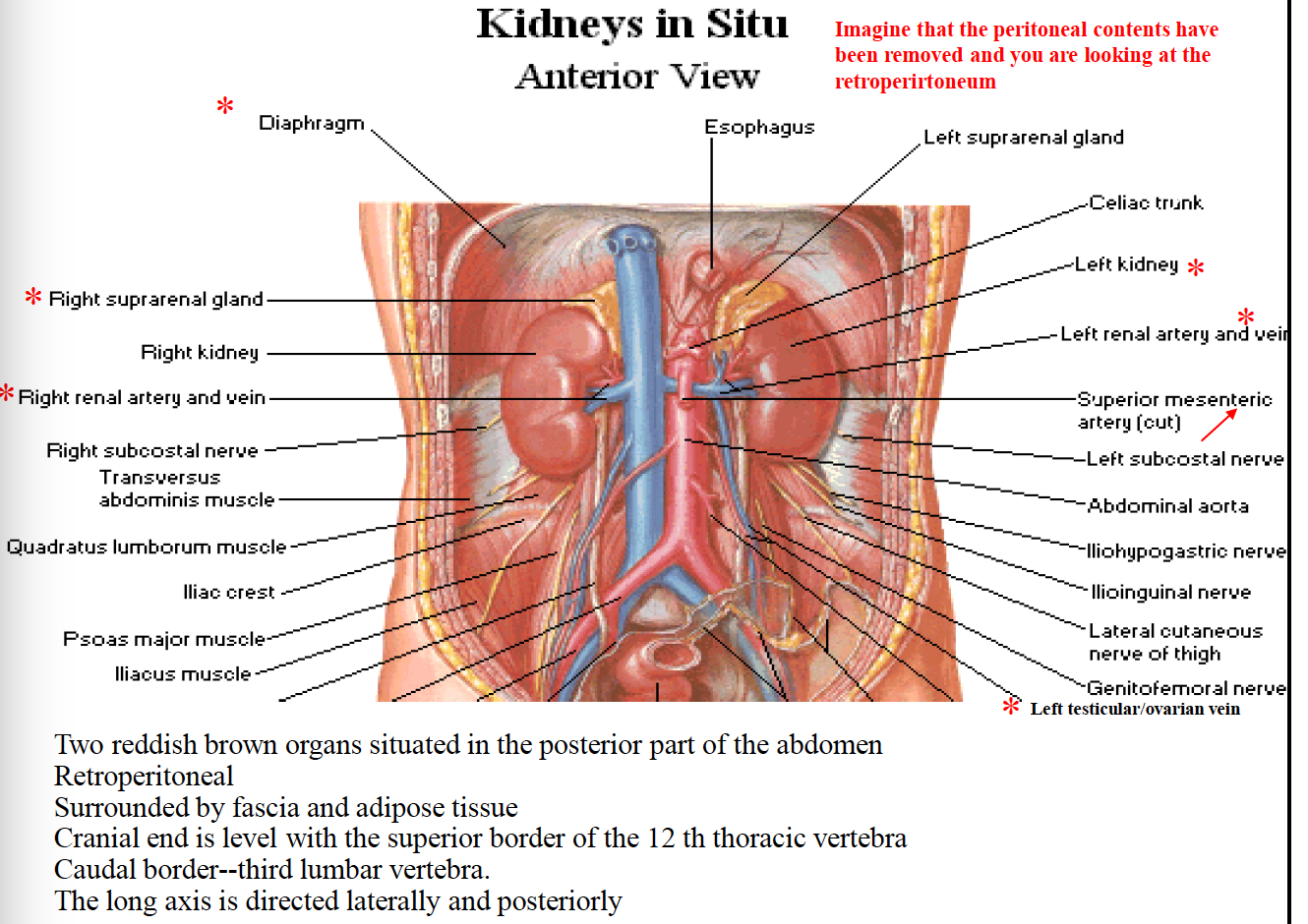
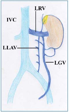
 Note Left Gonadal Veins Drains Into Left Renal Vein While
 Jcdr Anatomical Variants Gonadal Vein Morphology
 Renal Vein An Overview Sciencedirect Topics
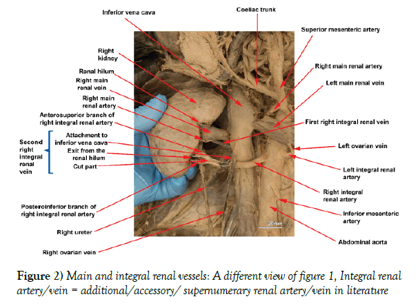 Integral Segmental Bi Lateral Renal Arteries And Unilateral

 Nutcracker Syndrome Servier Phlebolymphologyservier
 Renal Artery And Vein In Situ Renal Vasculature
 Open Access Journal Of Surgery Oajs Juniper Publishers
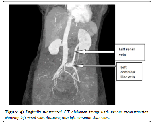
 Figure 1 From The Nutcracker Syndrome Semantic Scholar
 An Unusual Case Of Left Venous Renal Entrapment Syndrome A
 Anatomy Of Major Abdominal Veins Inferior Vena Cava
 Anatomy Of The Renal System Diagram Quizlet
 The Anatomy Of The Domestic Animals Veterinary Anatomy
![]() Kidneys Anatomy Function And Internal Structure Kenhub
Kidneys Anatomy Function And Internal Structure Kenhub
 Interlobar Arteries An Overview Sciencedirect Topics
 Which Of The Following May Have Least Amount Of Urea A Vena
 Renal Vasculature Radiology Key
 Schematic Diagram Showing Embryogenesis Of Inferior Vena
 Anomalous Adrenal Vein Anatomy Complicating The Evaluation
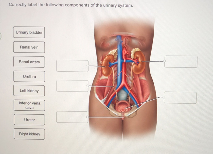 Solved Correctly Label The Following Components Of The Ur
 Urinary System Radiology Key
 Table Table 3 Definitions Of Tnm Stage Iiia Pdq Cancer
Surgical Anatomy Of The Retroperitoneum Abdominal Key
Belum ada Komentar untuk "Renal Vein Anatomy"
Posting Komentar