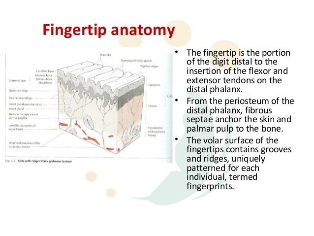
Fingertip Anatomy
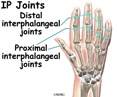
Distal interphalangeal dip proximal interphalangeal pip and metacarpophalangeal mcp. Finger fractures may account for up to 10 of all bone fractures.
 Surface Anatomy Hand Surgery Source
Ask a doctor online now.
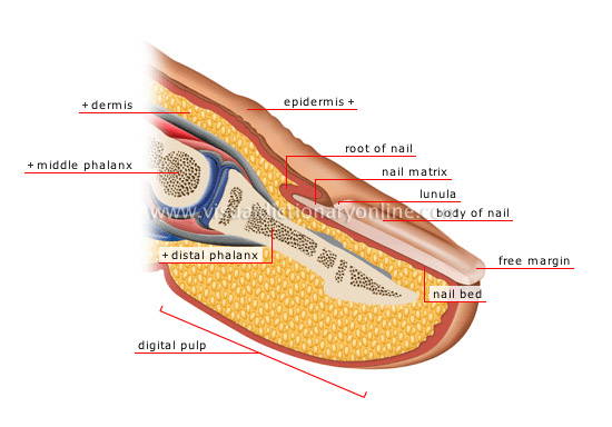
Fingertip anatomy. There are two interphalangeal joints ip joints on each finger. The fingertip wound should be assessed for tissue loss. You can also damage the nerve endings in the fingertips.
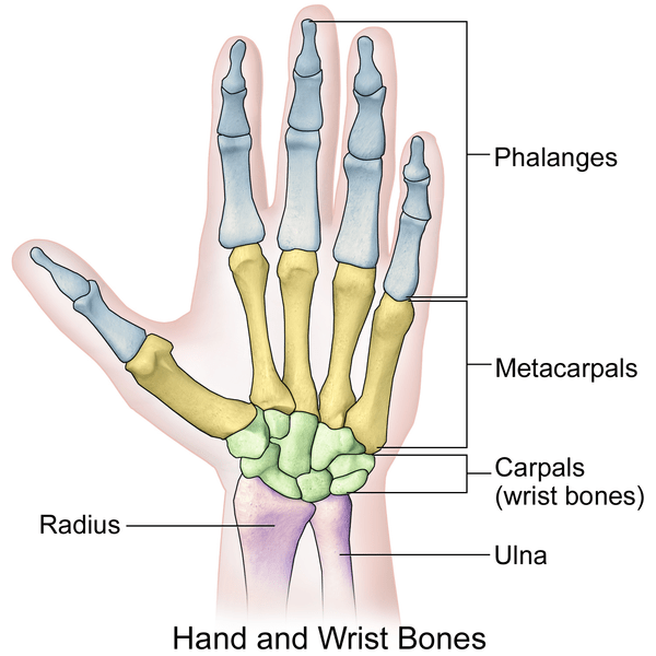
With high resolution 3 t mri the complex anatomy of the fingers can be imaged in exquisite detail to provide an accurate diagnosis of clinically important ligament and tendon injuries. The index middle ring and fifth digits have proximal middle and distal phalanges and three hinged joints. Ligaments connect finger bones and help keep them in place.
Anatomy of the fingers finger bones. Each finger has 3 phalanges bones and 3 hinged joints. The finger bones are known as phalanges singular phalanx.
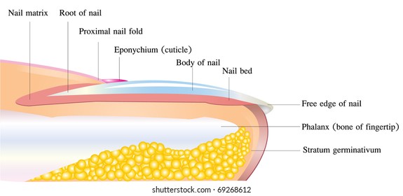
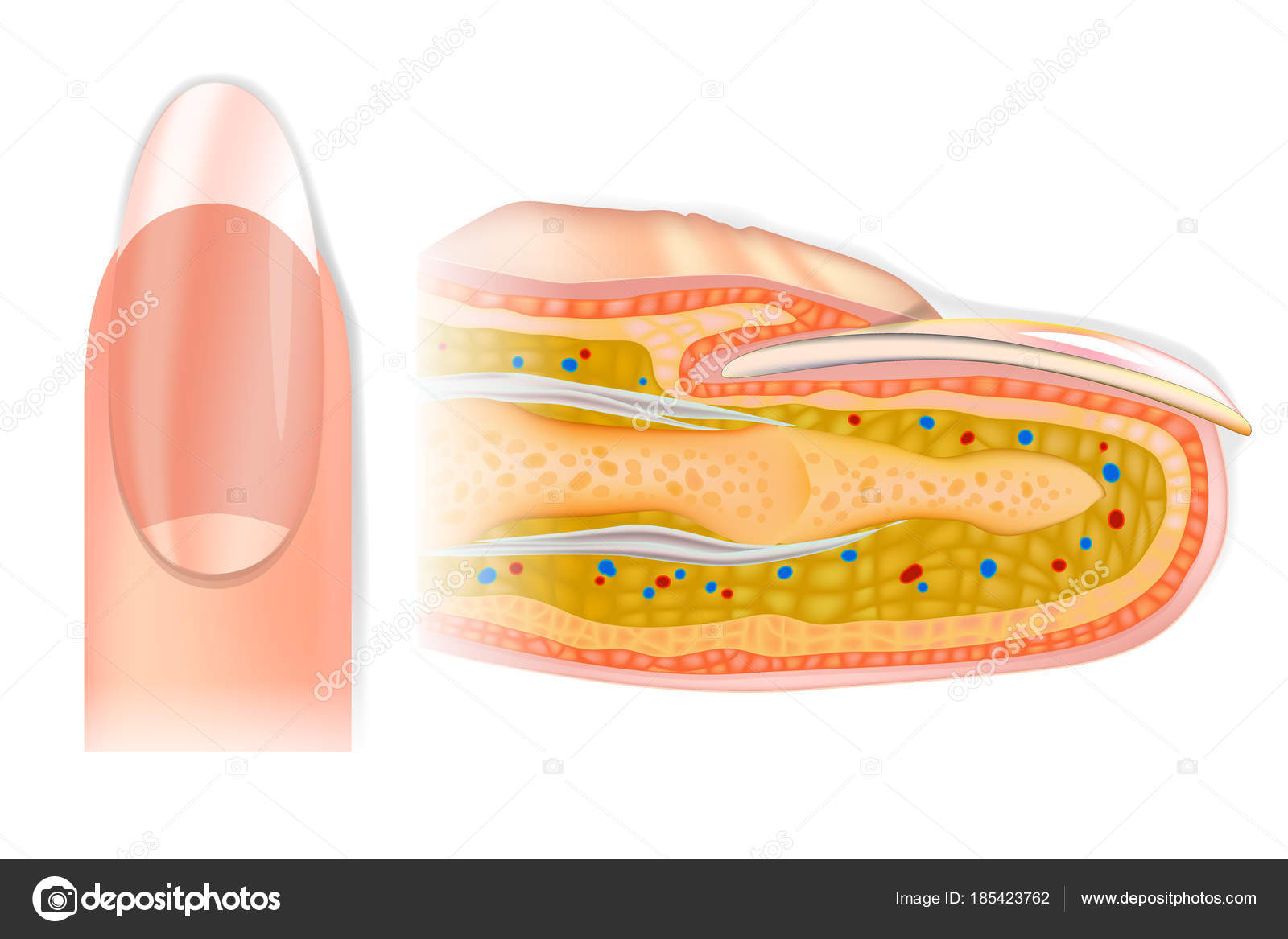
A fingertip injury can result in damage to the skin bone nailbed tendons and the pulp the padded area of the fingertip see figure 1. Like all skin it is made of two types of tissues. Bone support for nail growth.
The proximate phalange provides the base of the finger which connects to the intermediate phalange via the. Nerves of the fingers. Tendons connect muscles to bones.
If there is minimal tissue loss the wound can be debrided and closed primarily. Fingers have a complex anatomy. Injury to the finger with variable involvement of soft tissue bone and tendon.
If the fingertip wound cannot be repaired primarily no bone. The deeper dermis the living tissue which includes capillaries and glands and the epidermis the layer just beneath the nail plate which moves toward the finger tip with the plate. Goals of treatment sensate tip.
Prognosis improper treatment may result in stiffness and long term functional loss. The nail bed is the skin beneath the nail plate. Basic anatomy of the finger.
The finger is comprised of three phalanges extending from the hands second metacarpal. Fingers are easily injured and broken fingers are some of the most common traumatic injuries seen in an emergency room. Nerves send signals from the brain to the.
The anatomy of the finger is complex but a basic knowledge is necessary to properly treat acute injuries. The thumb has two of each.
 Finger Anatomy Images Stock Photos Vectors Shutterstock
 Hand Anatomy Eorthopod Com
 Preview
Hand Anatomy And Function Bone And Spine
 Finger Anatomy Picture Image On Medicinenet Com
 Fingertip Injuries Singhealth
 Finger Anatomy Archives Md Direct
Carpal Tunnel Syndrome Symptoms And Treatment Orthoinfo
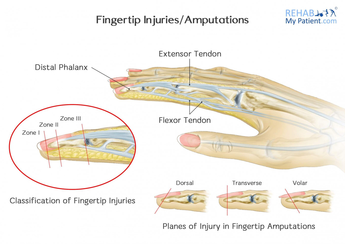 Fingertip Injuries Amputations Rehab My Patient
 Your Finger Joint Pain Is Probably Caused By Arthritis
 Nail Bed Injury Hand Orthobullets
 Hand Anatomy Eorthopod Com

 Acute Finger Injuries Part I Tendons And Ligaments
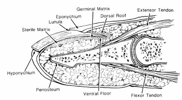
 Anatomy And Physiology Of The Fingertip Springerlink
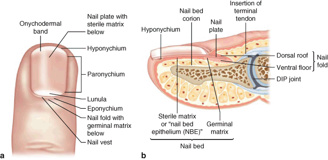
 Human Nail Finger Anatomy External View And The Sectional
 Extensor Tendon Lacerations Background History Of The
 Hand And Wrist Injuries Part I Nonemergent Evaluation
 Fingertip Recon
 Finger Amputation An Overview Sciencedirect Topics
 Finger Cross Section Anatomy Human Cross Section Finger
 Dupuytren Anatomy Dupuytren Research Group
 Figure 1 From Soft Tissue Injuries Of The Fingertip Methods
 Fingertip Amputations Finger Flaps Hand Orthobullets
 Anatomy And Physiology Of The Fingertip Springerlink
 Anatomy And Physiology Of The Fingertip Springerlink
Belum ada Komentar untuk "Fingertip Anatomy"
Posting Komentar