Cervical Vertebral Body Anatomy
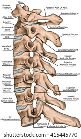
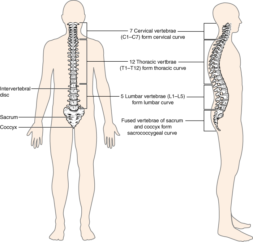
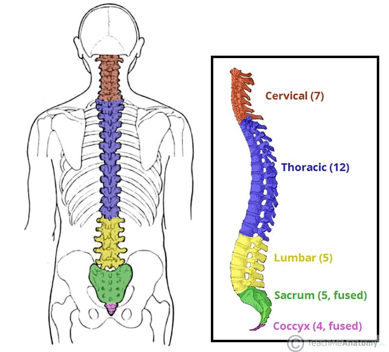
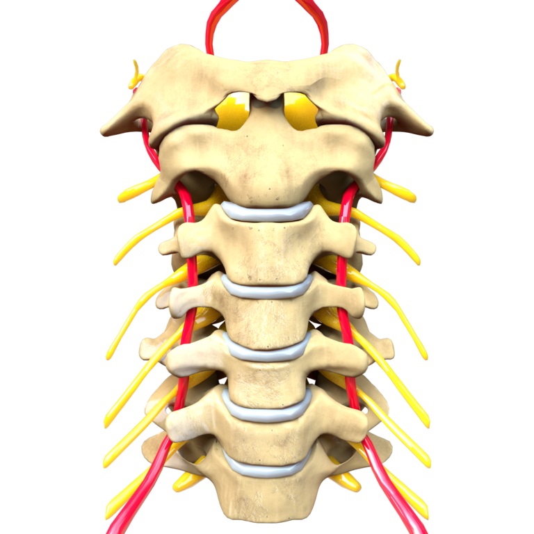
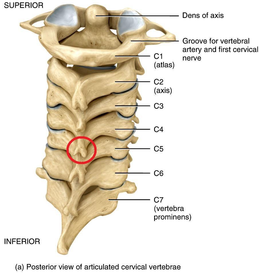
There are seven cervical vertebrae in the human body. Gross anatomy upper cervical spine.
 Cervical Vertebra An Overview Sciencedirect Topics
It consists of seven distinct vertebrae two of which are given unique names.

Cervical vertebral body anatomy. The twelve thoracic vertebrae are medium sized. Classifications of vertebrae cervical vertebrae. The cervical spine consists of seven vertebrae which are the smallest and uppermost in location within the spinal column.
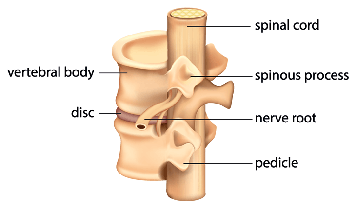
The top of the cervical spine connects to the skull and the bottom connects to the upper back at about shoulder level. Together the vertebrae support the skull move the spine and protect the spinal cord a bundle of nerves connected to the brain. An overhead view of the fourth cervical vertebra is typically shaped as the other vertebrae in thoracic and lumbar spine.
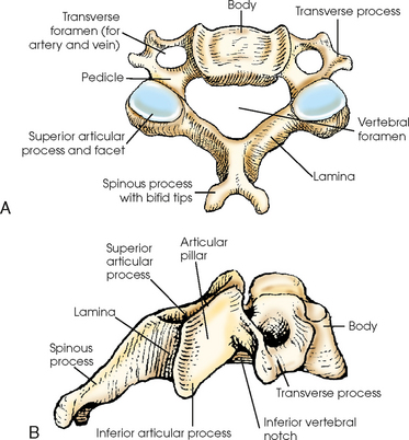
The upper surface is concave transversely and presents a projecting lip posterolaterally on. The illustration below is an overhead view of the fourth c4 cervical vertebra. The cervical spine is the most superior portion of the vertebral column lying between the cranium and the thoracic vertebrae.
To truly understand how indispensable this part of your back is we must point out that the complex nature of its composition makes it prone to various stresses forces conditions and ailments. Anatomy of the cervical spine. In this article we shall look at the anatomy of the cervical vertebrae.
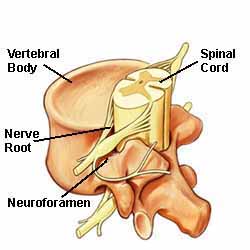
The vertebral foramen is a large opening in the center of the vertebra that provides space for the spinal cord and its meninges as they pass through the neck. The cervical spine starts at the bottom of the skull and wanders down through the seven vertebral bones mentioned earlier eventually connecting to the thoracic spine upper back. All seven cervical vertebrae are numbered.
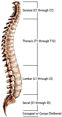
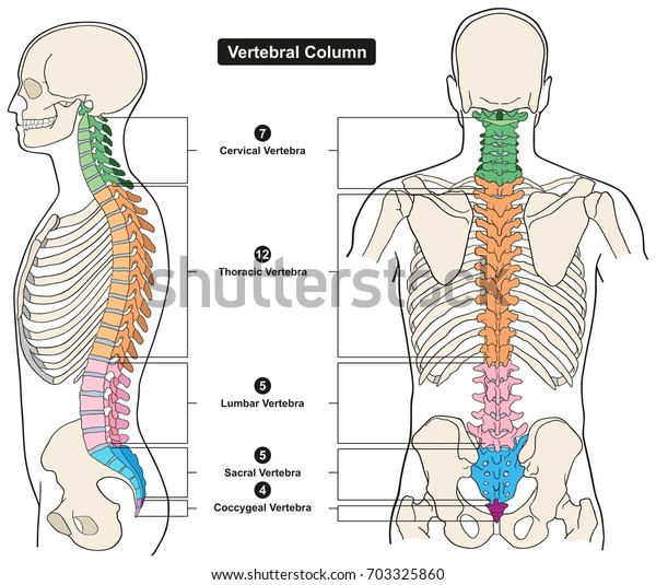
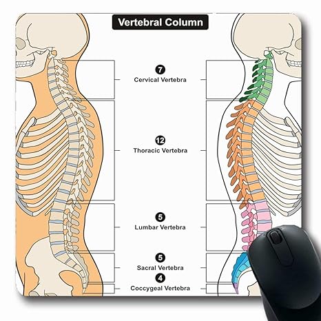
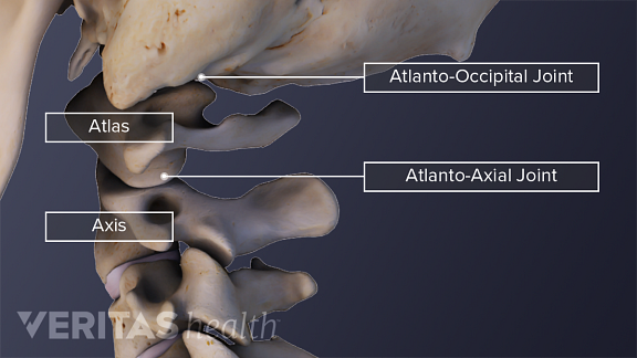
There are five lumbar vertebrae in most humans. The upper cervical spine consists of the atlas c1 and the axis c2. The cervical spine has 7 stacked bones called vertebrae labeled c1 through c7.
The lower surface is. These vertebrae have a box shaped body vertebral body. Vertebra of the neck.
The facet joints in the cervical spine are diarthrodial synovial joints. Vertebral body the bodies of these four vertebrae are small and transverse diameter is greater than. The first cervical vertebrae c1 is known as the atlas.
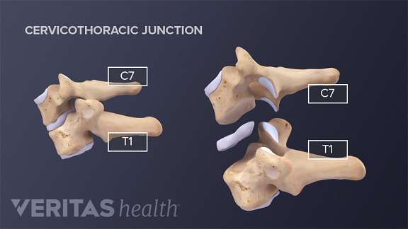
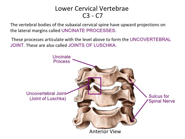
The third through sixth cervical vertebrae c3 c6 are different from the atlas and axis. The 5 cervical vertebrae that make up the lower cervical spine c3 c7. As viewed from the side the cervical spine forms a lordotic curve by gently curving toward the front of the body and then back.
Cervical spine anatomy video. The anterior and posterior surfaces are flattened and of equal depth. The sacrum is a collection of five.
Each cervical vertebra consists of a thin ring of bone or vertebral arch surrounding the vertebral and transverse foramina. The second cervical vertebrae c2 is known as the axis.
 The Main Weight Of The Body Is Carried By The Vertebral
 Spinal Cord Column Spinal Cord Injury Information Pages
 Spine Anatomy About The Spine Virginia Spine Institute
 Cervical Vertebrae Images Stock Photos Vectors Shutterstock
 Spine Anatomy Princeton Brain Spine And Sports Medicine
 Vertebral Column Human Body Anatomy Infograpic Education
 Anatomy Of Vertebra Column Ashiq
 Typical Cervical Vertebrae And C7 The Art Of Medicine
:background_color(FFFFFF):format(jpeg)/images/library/9053/cervical-spine-bones-and-ligaments_english__1_.jpg) Cervical Spine Anatomy Ligaments Nerves And Injury Kenhub
Cervical Spine Anatomy Ligaments Nerves And Injury Kenhub
 7 3 The Vertebral Column Anatomy And Physiology
 The Vertebral Column Joints Vertebrae Vertebral Structure
 Vertebral Column Radiology Key
 Explaining Spinal Disorders Cervical Stenosis
 Typical Cervical Vertebrae And C7 The Art Of Medicine
 Cervical Vertebrae Definition Function Structure
 Cervical Spine Anatomy Clinical Significances Anatomy Info
 Amazon Com Emvency Mouse Pads Vertebral Column Of Human
 Amazon Com Rwyzpad Mousepads Cervical Skull Vertebral
 Typical Cervical Vertebrae And C7 The Art Of Medicine


Belum ada Komentar untuk "Cervical Vertebral Body Anatomy"
Posting Komentar