Liver Anatomy Dog
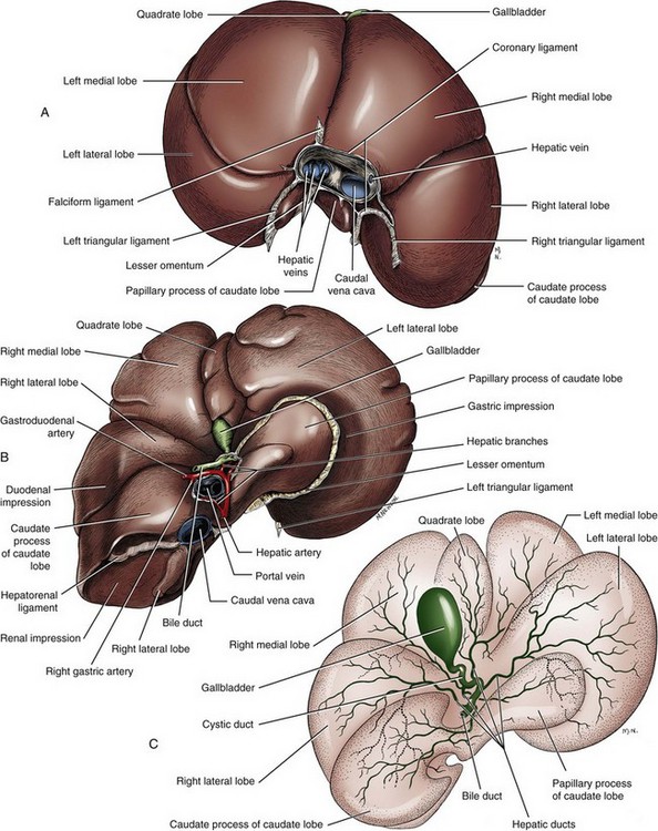
The caudate lobe extends dorsally and caudally ending with the renal fossa which contains the cranial pole of the right kidney. Complete obstruction of the hepatic artery is fatal.
 Canine Anatomy Illustrations Lovetoknow
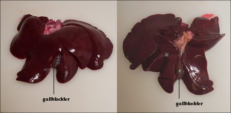
In dogs the livers left lobe medial sublobe quadrate lobe and right lobe medial sublobe encircle the gallbladder.

Liver anatomy dog. Alt ast ggt albumin and bilirubin are important values on the chemistry profile that are often abnormal in dogs that have liver disease. Fortunately the normal liver has an incredible regenerative capacity. Canine liver and anatomy canine liver disease is among the top five leading causes of non accidental death in dogs and as such should be taken seriously.
Other common symptoms of liver disease are gastrointestinal signs such as decreased appetite vomiting and diarrhea weight loss increased drinking and urination and changes in stool color. These further extend to the heel bone known as tarsus the paw bone known as metatarsus and the toe bone phalange. The liver is almost entirely intra thoracic.
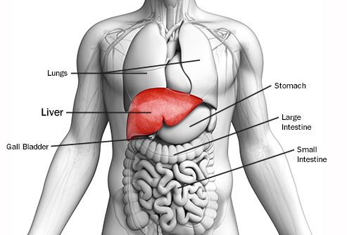
It is so far forward that it lays up against the diaphragm the muscle that aids in breathing in mammals birds and reptiles do not have a diaphragmthis autopsy picture of a cat shows the gallbladder in green with several lobes of the liver laying right up against the diaphragm towards the top of the. Signs of liver disease in dogs. The liver is responsible for a number of essential bodily functions and if it is compromised in any way your dogs overall health is in jeopardy.
The liver is a multi lobed organ that is located at the most forward part of the abdomen. The liver is contained entirely within the rib cage to the right of the midline. An enlarged caudate process contacts the right kidney.
Dogs may develop fluid retention in the abdomen commonly referred to as ascites. Abnormalities in the metabolism of glucose and fat in diabetic dogs cause an accumulation of fat in the liver that may eventually result in liver dysfunction. It is less lobated.
Both the left and right lobes are subdivided. Liver disease in dogs is diagnosed through history physical examination complete blood count bile acid testing urinalysis radiographs x ray abdominal ultrasound and biopsy. Steroid hepatopathy is a condition in which there is excessive glycogen deposition in liver cells due to high circulating levels of steroids in the blood.
Vomiting loss of appetite or diarrhea. In experimental studies normal dogs tolerated acute removal of 65 to 70 of total liver volume but they did not tolerate 84 removal154 204 and approximately 28 of dogs survived at least 7 days after 80 hepatectomy. The rear legs of the dog begin with the femur bone which extends to a pair of bones known as the tibia and the fibula.
A liver sick dog may also present with.
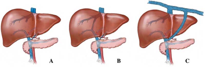 File Dog Liver Portosystemic Shunts Jpg Embryology
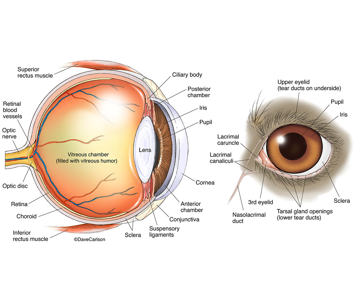 Dog Eye Anatomy Carlson Stock Art

 Labeled Atlas Of Anatomy Illustrations Of The Dog
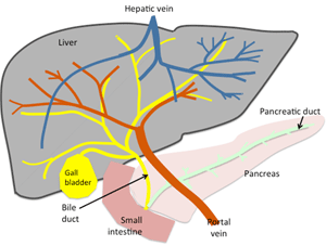
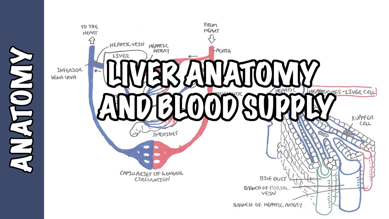 Liver Anatomy And Blood Supply
 Canine Anatomy Learn About Animal Anatomy
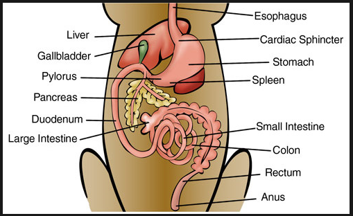
Liver Pancreas Spleen
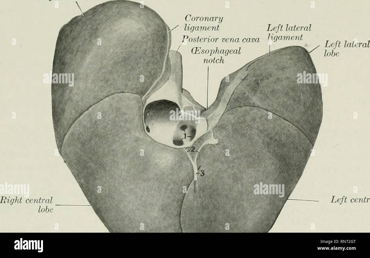 The Anatomy Of The Domestic Animals Veterinary Anatomy Fig
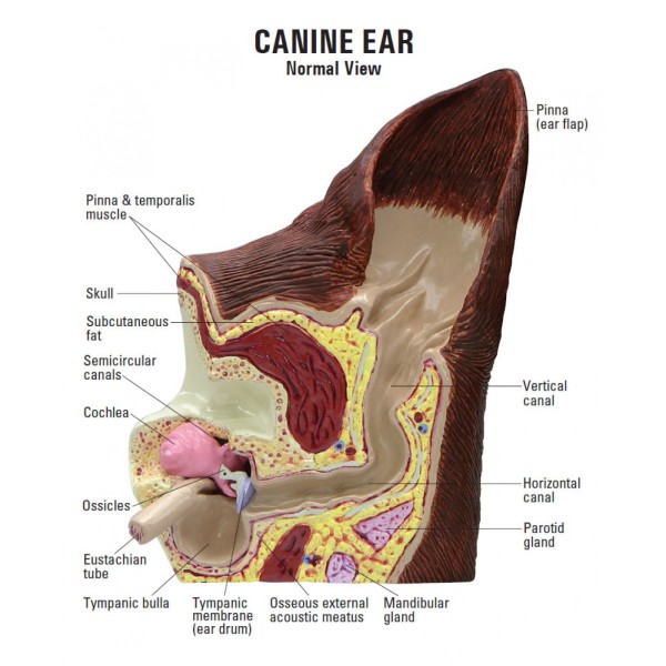 Ear Disease
 Abdomen And Pelvis Anatomy Of The Dog On Ct
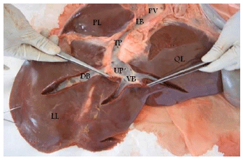
 Liver Lobes Anatomy Liver Anatomy Bile Duct Dog Anatomy
 Liver Dis Ease In Your Pet The Whole Dog
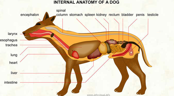 Internal Anatomy Of A Dog Visual Dictionary
Pancreatitis In Dogs Vca Animal Hospital
 Canine Anatomy Illustrations Lovetoknow

Liver Pancreas Spleen
 Visual Guide To Liver Cancer
 Anatomy Of A Dog
 Dog Muscular Skeletal Nerves Canine Anatomy Poster 18 X 24 Veterinary Chart
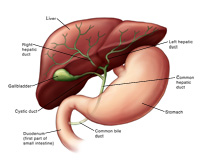
 Liver And Biliary System Veterian Key
Belum ada Komentar untuk "Liver Anatomy Dog"
Posting Komentar