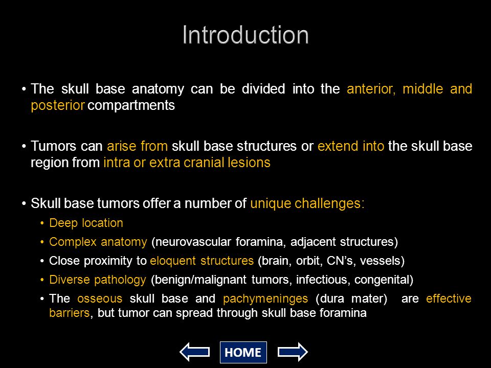
Ct Skull Base Anatomy
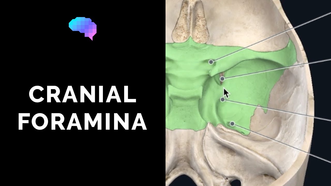
Navigating the skull base. Cranial foramina for further details.
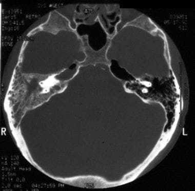
 Emdocs Net Emergency Medicine Educationbasilar Skull
An interactive program for learning skull base anatomy welcome to navigating the skull base.

Ct skull base anatomy. Basic skull base anatomy. You can however go directly to any section using the links below. To begin click on introduction.
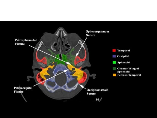
Skull ct anatomy the sagittal suture is the line where the right and left parietal bone are in contact. Foramina of the skull base and the structures that pass through them. Navigating the skull base identify the petro occipital fissure to navigate the major structures of the skull base.
Ct anatomy of skull base. Basic anatomy review the bones sutures and fissures that comprise the skull base. Detailed anatomy enter this module for a more detailed review of skull base anatomy.
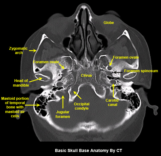
The module interface is meant to mimic a radiology workstation with adjacent image scrolling via arrow keys and or mouse wheel button. For lesions that are completely intraosseous the mass may only be appreciated on mri figure 1. The skull base can be evaluated by computed tomography ct which will demonstrate the bony structures of the skull base with its foramina and fissures for vessels and cranial nerves the temporal bone and sinonasal cavities.
Given that the file is large loading may take a few minutes. Ct is superior to mri for subtle cortical bone changes but marrow processes are best characterized with non gadolinium gd contrast enhanced t1 mri. Ct is the modality of choice in defining the bony anatomy of the skull base and to depict the thin cortical margins of skull base neurovascular foramina.
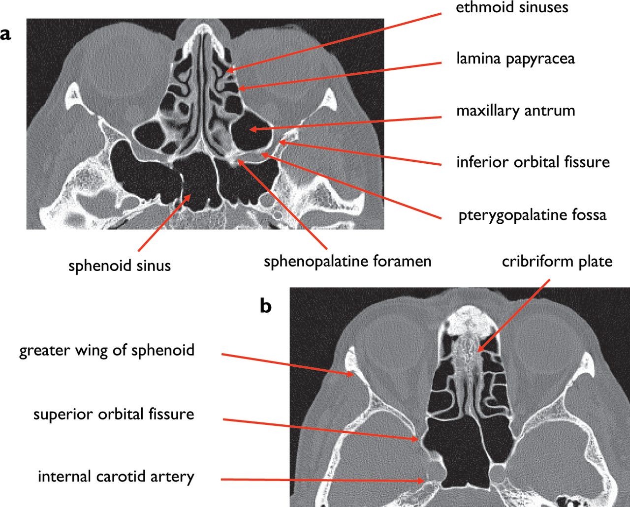
2 superior orbital fissure. A axial three dimensional reconstructed ct image with color coded overlay shows the skull base sections. Blue central skull base csb purple posterior skull base teal anterior skull base asb.
Ct anatomy of skull base. Blue temporal bones fuchsia nasal bones green ethmoid bone light pink vomer purple occipital bones teal sphenoid bone yellow zygomatic bones. Ct is more sensitive in detecting fibro osseous skull base lesions calcification and sclerosis.
A noncontrast sinus or skull base ct which covers the mastoids temporal bone and entire skull base is recommended and intravenous contrast is not usually necessary as the mri will provide soft tissue detail. 4 mastoid air cells. B axial ct image with color coded overlay shows the skull base bones.
To load the skull base ct anatomy module in a new window click on its image above. Click the next link at the bottom of each page to step through each section. The coronal suture is the line where the parietal bone frontal bone and are in contact.
3 anterior clinoid process. The base of the skull is perforated by numerous foramina which allow vessels and nerves to pass through the base of the skull allowing the intracranial cavity to communicate with the rest of the body see main article. 5 internal occipital protuberance.
 Normal Anatomy Of The Base Of The Skull Orbit Pituitary
 Normal Anatomy Of The Base Of The Skull Orbit Pituitary
 Anatomy And Pathology Of The Skull Base Ct And Mri Imaging
 3d Printed Skull Base Generated From Ct Scan Data Accurately
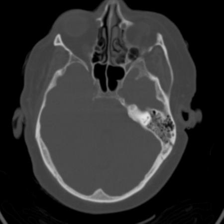
 Fibrous Dysplasia Of The Skull Base Axial Ct Of The Head In
 Sagittal Ct Bone Window Of Skull Base Open I
 Imaging Of Skull Base Tumors Henry Ford Health System Ppt
 Ct Scan Tips Protocols Ct Skull Base Anatomy
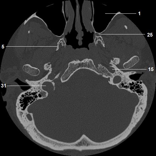 Temporal Bone And Skull Base Anatomy Ct
North Jersey Brain Spine Center Computerized Tomography
Anatomy And Pathology Of The Skull Base Ct And Mri Imaging
 Brain And Face Ct Interactive Anatomy Atlas
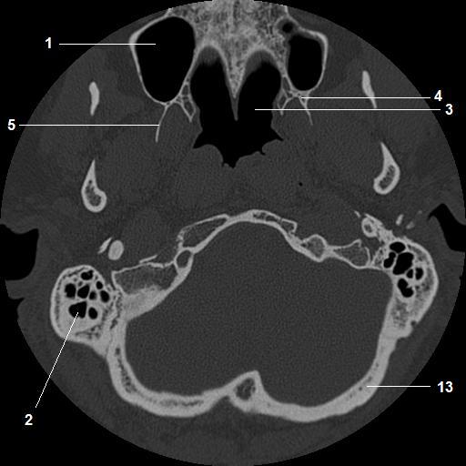 Ct Temporal Bone Skull Base Anatomy Axial
 Mr Ct And Plain Film Imaging Of The Developing Skull Base
 Skull Base Intraosseous Lipoma Radiology Case
 Imaging Of Skull Base Pictorial Essay Raut Aa Naphade Ps
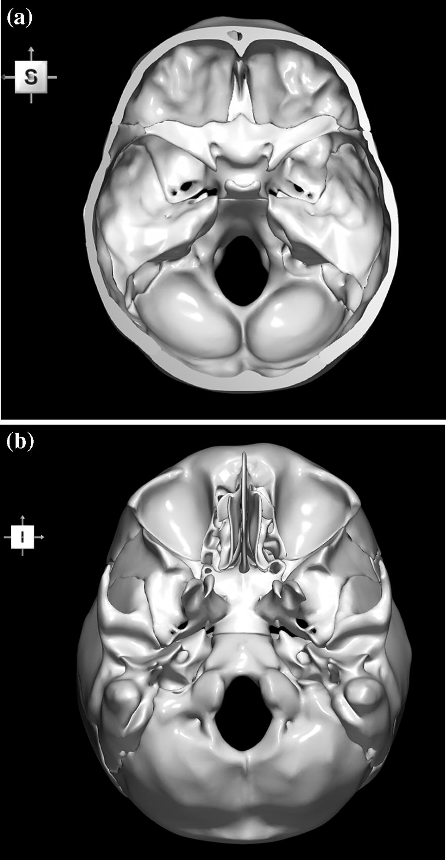 A 3d Stereotactic Atlas Of The Adult Human Skull Base
 A 3d Stereotactic Atlas Of The Adult Human Skull Base
 Skull Base Tumors Uci Head And Neck Surgery Uci Ent
 Imaging Of Skull Base Pictorial Essay Raut Aa Naphade Ps
 Imaging Of Skull Base Pictorial Essay Raut Aa Naphade Ps
 Skull Base Petrous Apex Tumors Background History Of The
 Foramen Of The Skull Cranial Nerves 3d Anatomy Tutorial
![]() Superior View Of The Base Of The Skull Anatomy Kenhub
Superior View Of The Base Of The Skull Anatomy Kenhub
 Skull Base Anatomy Axial Plane Infant Skull Base Ct
 Untitled Document
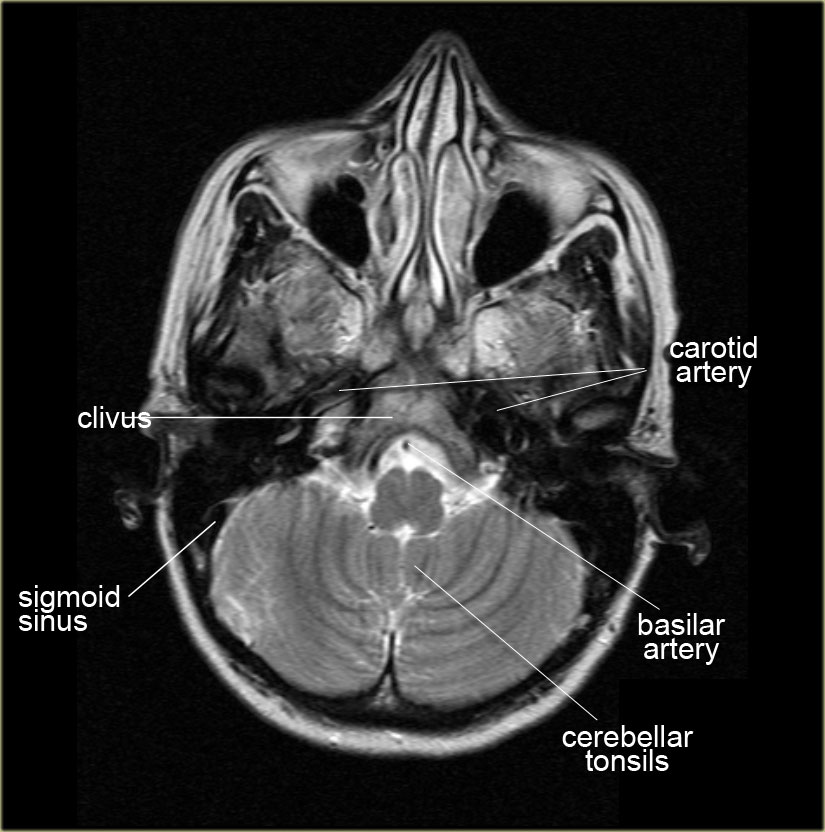 The Radiology Assistant Brain Anatomy
 Ct Scan Tips Protocols Ct Skull Base Anatomy
 A Review Of Basilar Skull Fractures Emra
 Brain And Face Ct Interactive Anatomy Atlas
 Skull Base Tumors Background History Of The Procedure Problem
Ct Scan Of Brain And Base Of Skull Stock Photo Download
 Imaging In Skull Base


Belum ada Komentar untuk "Ct Skull Base Anatomy"
Posting Komentar