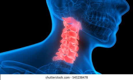
Cervical Spine Mri Anatomy
Cervical radiculopathy workup mri the american college of radiology recommends routine mri as the most appropriate imaging study in patients with chronic neck pain who have neurologic signs or symptoms but normal radiographs. This anatomy section promotes the use of the terminologia anatomica the global standard for correct gross anatomical nomenclature.
 Facet Joint Syndrome From Diagnosis To Interventional
This photo gallery presents the anatomical structures found on cervical spine mri t2 weighted axial and sagittal views.

Cervical spine mri anatomy. 6 inferior endplate c2. The neck or cervical spine is the top part of the spine between. This module of human anatomy is dedicated to residents and students who wish to learn the basics of the anatomy of the cervical spine in mri on a 15 tesla device.
Mri has become the method of choice for imaging the neck to detect significant soft tissue pathology such as disc. The cervical spine has 7 stacked bones called vertebrae labeled c1 through c7. In particular an mri shows a cross section of your tissue and each cross section is so thin that a single mri actually creates hundreds of images of your neck.
The disc is the shock absorber in the front of. Mri of the cervical spine. 1 jugular vein and carotid artery.
This mri cervical spine sagittal cross sectional anatomy tool is absolutely free to use. Two nerve roots one on each side sprout from the cord at each level. Spinal discs in the neck.
Spinal anatomy encompasses the anatomy of all osseous and soft tissue structures of the spine the spinal cord and its supporting structures. A cervical spine mri is different from an x ray although both are imaging techniques. Whereas an x ray just shows your spine or neck bones an mri shows your soft tissues.
Cervical spine anatomy video. Nerves in the neck. As viewed from the side the cervical spine forms a lordotic curve by gently curving toward the front of the body and then back.
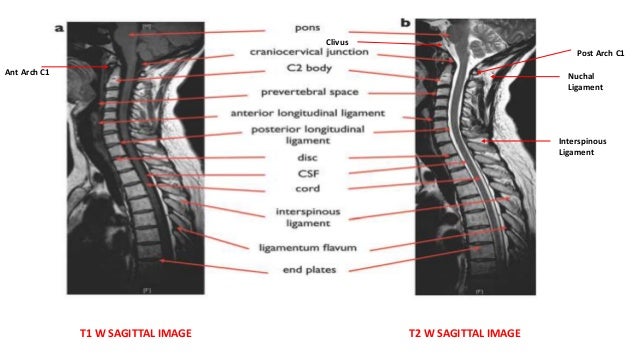
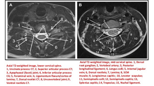
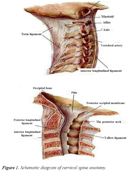
Anatomy of the cervical spine in magnetic resonance imaging mri cervical vertebrae spinal cord ligaments joints. Mri of the cervical spine axial t2 weighted image. Except for the first and the second cervical vertebrae the vertebrae share a similar structure including a vertebral body containing trabecular bone.
Use the mouse scroll wheel to move the images up and down alternatively use the tiny arrows on both side of the image to move the images. Anatomy of the cervical spine anatomy of the neck. The spine is composed of seven cervical twelve thoracic and five lumbar vertebrae as well as the fused sacrum and coccyx vertebral elements.
The top of the cervical spine connects to the skull and the bottom connects to the upper back at about shoulder level. What does a cervical spine mri show. This mri cervical spine c spine cross sectional anatomy tool is absolutely free to use.
Use the mouse scroll wheel to move the images up and down alternatively use the tiny arrows on both side of the image to move the imageson both side of the image to move the images.
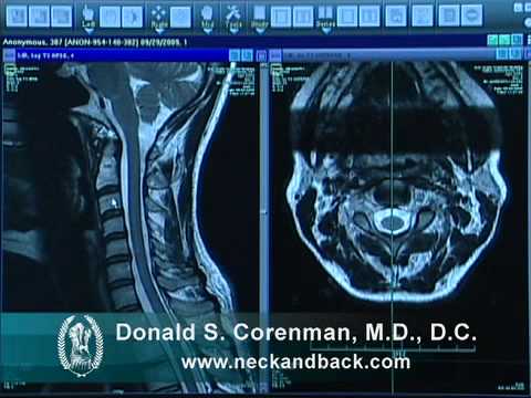 How To Read A Mri Of The Normal Cervical Spine Neck Colorado Spine Expert
 Mri Spine Anatomy Free Mri Axial Cervical Spine Anatomy
 Post Mva Cervical Spine Anatomy High Impact Visual
 Mri Spine Anatomy
 Facet Joint Syndrome From Diagnosis To Interventional
 Imaging Of The Cervical Spine
 Surgical Disorders Of The Cervical Spine Presentation And

 Radiology Images
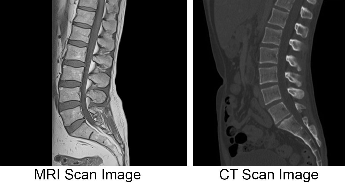 Mri Vs Ct Scan Diagnosing Spine Neck Injuries
 Surgical Disorders Of The Cervical Spine Presentation And
 Mri Of Cervical Spine In A 4 Year Old Patient With Morquio A
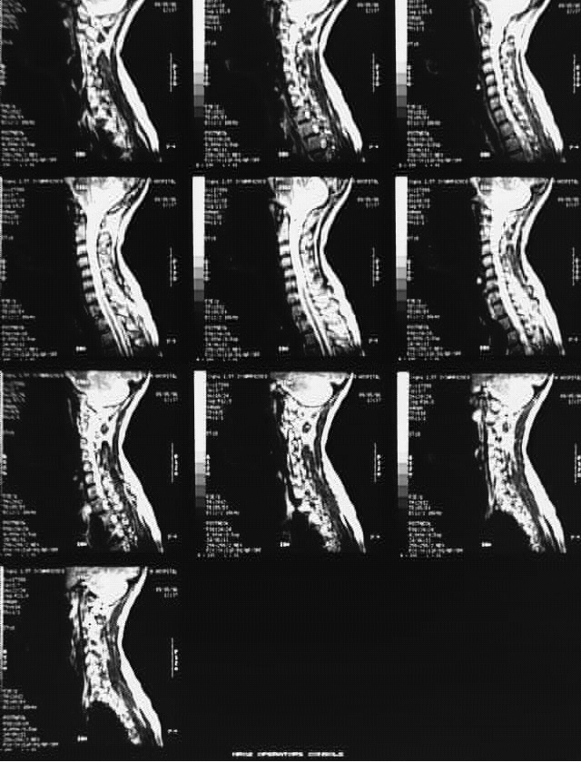
 Cervical Spine Mri

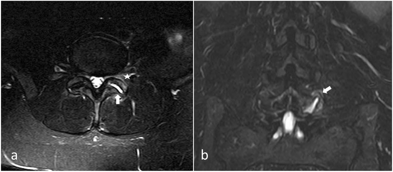
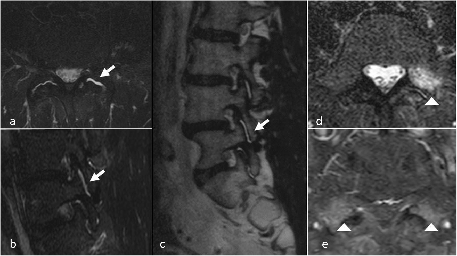
 Sonoanatomy Relevant For Ultrasound Guided Injections Of The
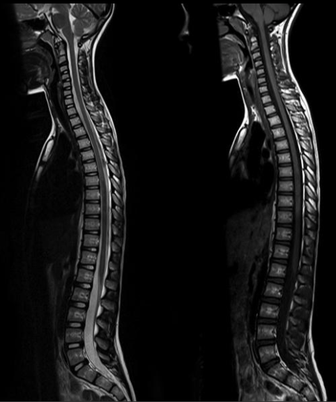
 Normal Cervical And Thoracic Spine Mri Radiology Case
Plos One An Initial Experience With The Use Of Whole Body
:max_bytes(150000):strip_icc()/GettyImages-1129624171-08e8d926acc944b697fc54acb2647660.jpg) Cervical Spine Curve And Injuries
Cervical Spine Curve And Injuries
 Cervical Spine Mri

 Mri Spine Anatomy

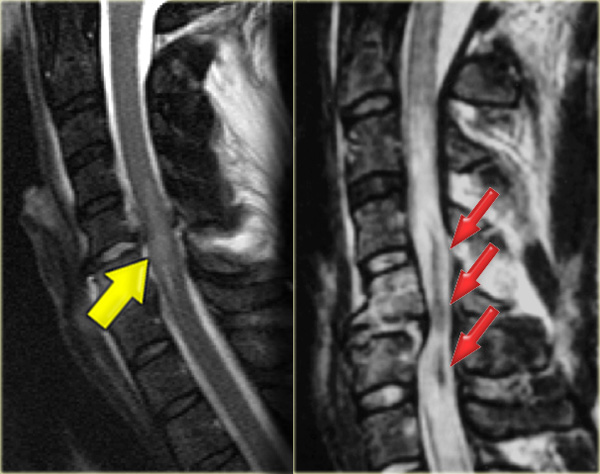
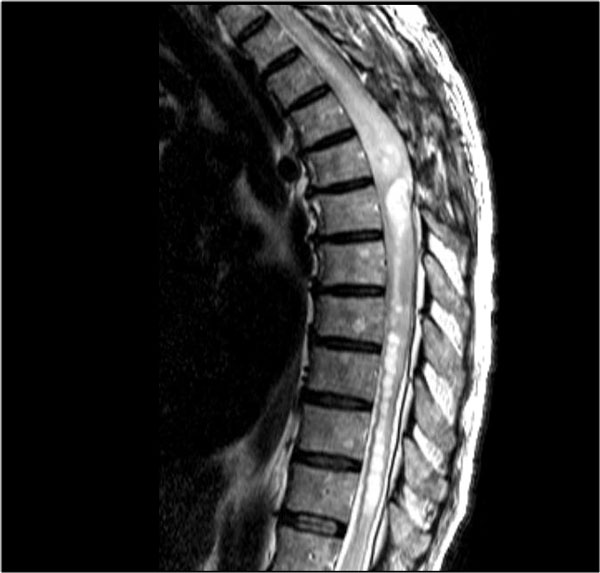 The Radiology Assistant Spine Myelopathy
 Cervical Spine Images Stock Photos Vectors Shutterstock
 Cervical Spine Mri
 The Radiology Assistant Spine Cervical Injury
 Normal Images Of Spine Joshua Li Md Phd
 Chiari Malformation
Belum ada Komentar untuk "Cervical Spine Mri Anatomy"
Posting Komentar