Pancreas And Spleen Anatomy
The tail of the pancreas abuts against the hilum of the spleen through which blood vessels lymphatics and autonomic nerves enter and leave the organ. The neck body and tail of the pancreas lie ventral to the splenic vein with the tail extending into the hilum of the spleen.
 The Pancreas Anatomy Duct System Vasculature
It is anterior to the spine the inferior vena cava and the aorta.

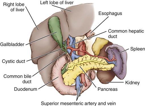
Pancreas and spleen anatomy. A sound appreciation of the topographical vascular and ductal anatomy of the pancreas is fundamental to the successful surgical management of pancreatic cancers congenital malformations of the pancreas and various surgical complications of acute pancreatitis. The spleen figure 5 anterior to the spleen lies the stomach. The longest part of the pancreas the body stretches across behind the stomach and the tail of the pancreas ends adjacent to the spleen.
The pancreas stretches from the inner curvature of the duodenum where the head surrounds two blood vessels the superior mesenteric artery and vein. The pancreas aids in digestion by producing enzymes that digest several types of nutrients including proteins fats carbohydrates and nucleic acid. Inferiorly the well named splenic flexure of the colon and medially the left kidney.
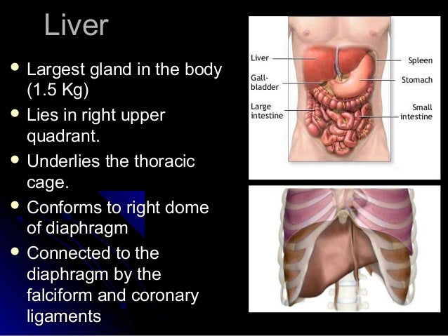
Play this quiz called liver pancreas and spleen anatomy and show off your skills. The spleen is the largest lymphoid organ in the body. Spleen located posteriorly and laterally.
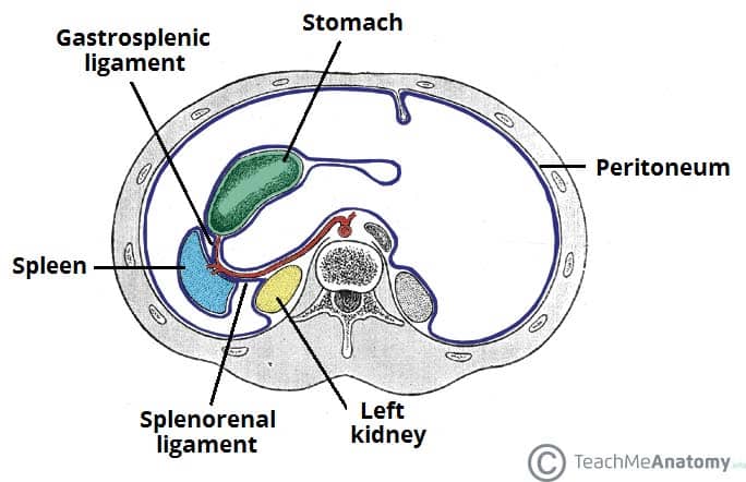
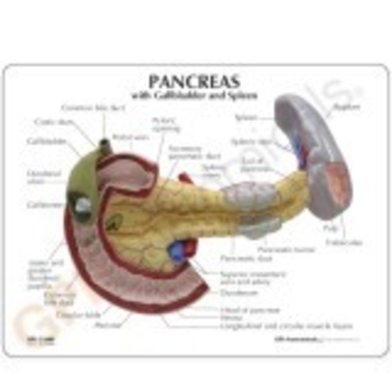
The somso pancreas spleen duodenum anatomy model is life size and made in somso plast. The pancreas is a wing shaped gland that extends from the duodenum the upper portion of the small intestine to the spleen. The lienorenal ligament is formed from peritoneum and connects the spleen to the tail of the pancreas.
This is a quiz called liver pancreas and spleen anatomy and was created by member ashpoprox login. The pancreas lies near several major vessels and significant landmarks in vascular anatomy. Bleeding is typically profuse due to the spleens thin capsule and soft parenchyma what is the cause of most cases of extrahepatic obstruction of biliary ducts leading to jaundice.
It serves both digestive and endocrine functions. Pancreatic tissue is best recognized by identification of the vessels around it. The aorta and inferior vena cava pass posteriorly to the head of the pancreas.
The pancreas has detail of the pancreatic duct shown up to its aperture and the duodenum is partly open.
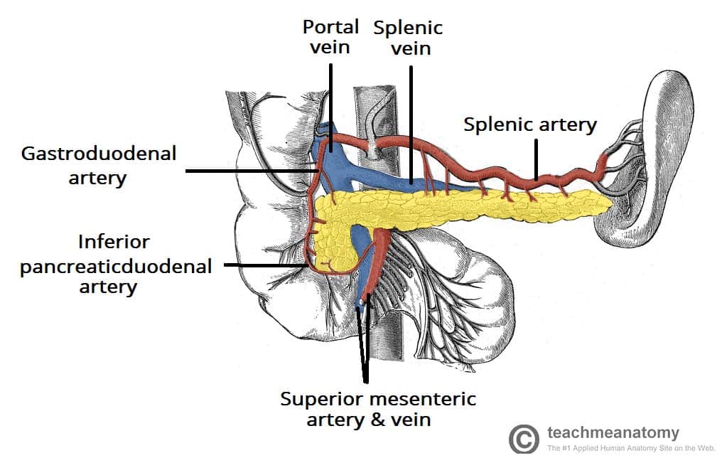
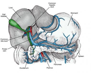
 Pancreaticosplenic Vessels Musculoskeletal Key
 Pancreas Gallbladder Spleen Anatomy Model
 Liver Gallbladder Pancreas And Spleen Labeled Female
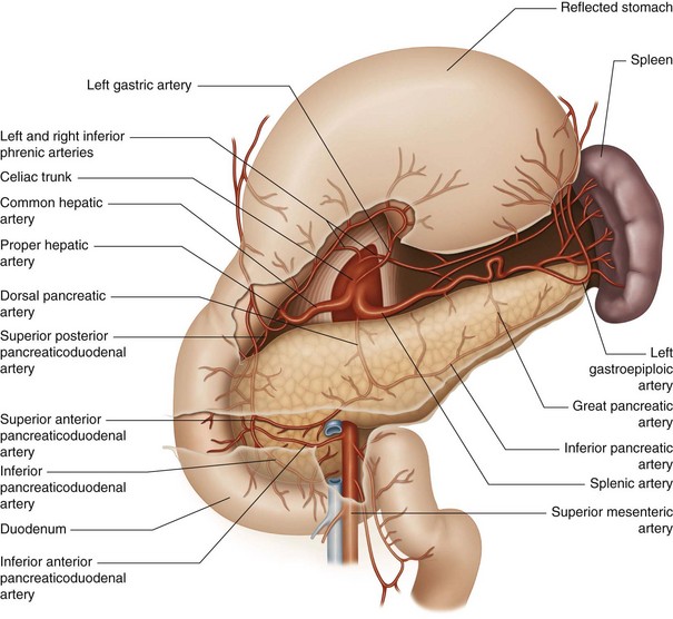
 Anatomy Of The Pancreas And The Spleen Sciencedirect
 Spleen Images Stock Photos Vectors Shutterstock
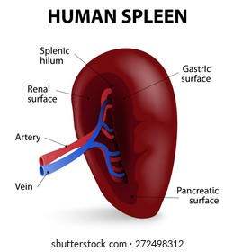
 The Spleen Position Structure Neurovasculature
Anatomy By411 The Spleen Pancreas And Gall Bladder
 Gallstones In The Gallbladder And Anatomy Of Duodenum Pancreas
 New Anatomical Diseased Pancreas Gallbladder Spleen
Splenic Arterial Interventions Anatomy Indications
 Pancreas And Spleen Stock Photos Pancreas And Spleen Stock
 Anatomical Model Pancreas Gb Spleen Cancer
 Liver Biliary System Pancreas And Spleen
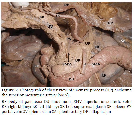 Histomorphological Study Of Unusual And Extensive Uncinate
 0146 00 Internal Organs
 Surgery Of The Liver Biliary Tract Pancreas And Spleen
 Pancreas Clinical Anatomy And Physiology
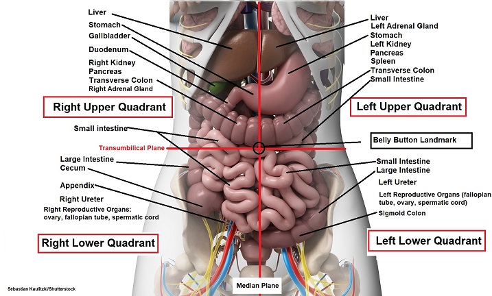 Four Abdominal Quadrants And Nine Abdominal Regions
 Anatomy And Physiology Ii Pancreas Spleen Duodenum Model
 Pancreatosplenectomy And Spleen Preserving Distal
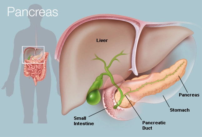
![]() Pancreas Anatomy Functions Blood Supply Innervation Kenhub
Pancreas Anatomy Functions Blood Supply Innervation Kenhub
 What Is The Pancreas Pancreatic Cancer Action Network
 Pancreas Human Anatomy Picture Function Conditions
Liver Pancreas Spleen
 Pancreas Wikipedia
 Pancreas Radiology Key
 Spleen And Surrounding Anatomy
 Lifesize Anatomical Human Pancreas Duodenum Spleen Model Anatomy Medical
 Arteries Of Stomach Duodenum Pancreas And Spleen
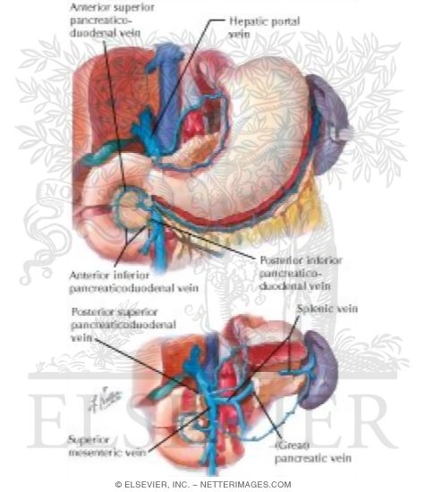 Veins Of Stomach Duodenum Pancreas And Spleen Venous


Belum ada Komentar untuk "Pancreas And Spleen Anatomy"
Posting Komentar