Anatomy Of Temporomandibular Joint
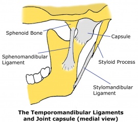
There are three extracapsular ligaments. The joint has a capsule and an articulating disc.
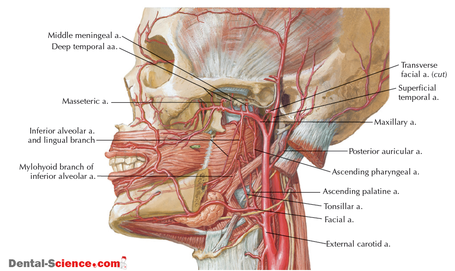
The tmj is supplied by three arteries.
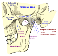
Anatomy of temporomandibular joint. The mandibular nerve. Temporomandibular joint disorder known more commonly as tmd occurs when there are problems with the muscles and jaws in the face. With the mandibular fossa glenoid fossa of the temporal bone.
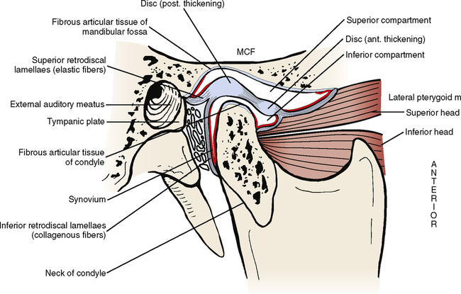
The joint is separated into a superior and an inferior compartment by. The temporomandibular joint articulating surfaces. The temporomandibular joint tmj has long since been established as a source of pathology 1 but did not become a central focus of research until the 1980s.
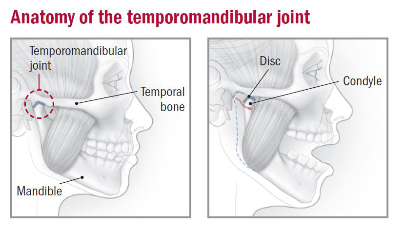
The temporomandibular joint consists of articulations between three surfaces. 2 collectively pathoanatomical dysfunctions of the tmj have been defined as temporomandibular disorders tmd. The temporomandibular joint tmj is an atypical synovial joint located between the condylar process of the mandible and the mandibular fossa and articular eminence of the temporal bone.
The two minor ligaments the stylomandibular and sphenomandibular ligaments are accessory and are not directly attached to. Currently both dentists and physical therapists provide patient services for tmd. The temporomandibular joint tmj or jaw joint is a bi arthroidal hinge joint that allows the complex movements necessary for eating swallowing talking and yawning.
The anatomy of the temporomandibular joint the anatomy of the temporomandibular joint by david terfera shereen jegtvig the temporomandibular joint is a modified hinge type of synovial joint made up of the condylar process of the mandible and the mandibular fossa of the temporal bone. An outer oblique portion oop. Anatomy and physiology of temporomandibular joint temporomandibular joint tmj.
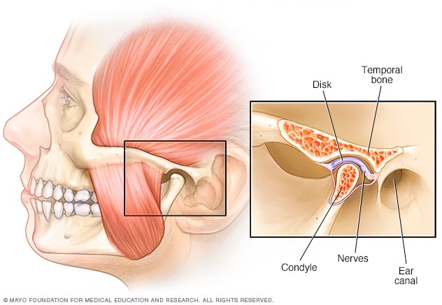
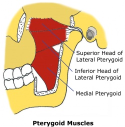
Movements at this joint are produced by the muscles of mastication. Ligaments the major ligament the temporomandibular ligament is actually the thickened lateral portion of the capsule and it has two parts. It is the articulation of the condyle of the mandible and the inter articular disc.
It is divided into a superior discotemporal space and inferior discomandibular space by the tmj disc or meniscus. The temporomandibular joint tmj is the joint that connects the jaw to the temporal bones of the skull. Dysfunction of the tmj can cause severe pain and lifestyle limitation.
The joint capsule originates from the border of the mandibular fossa.
 Detailed Anatomy Of The Temporomandibular Joint Springerlink
 Tmj Temporomandibular Joint Pain Lake Zurich Il Patch
Temporomandibular Joint Dysfunction Harvard Health
 Temporomandibular Joint Mayo Clinic
 Dentistry Lectures For Mfds Mjdf Nbde Ore Anatomy Of
 Tmj Therapy
 Stop Suffering From Tmj Temporomandibular Joint Pain
 Tmj Anatomy Physiopedia
 29 Temporomandibular Joint Pocket Dentistry
 Tmj Anatomy Physiopedia
_Anatomy.png)
 The Temporomandibular Joint Musculoskeletal Key

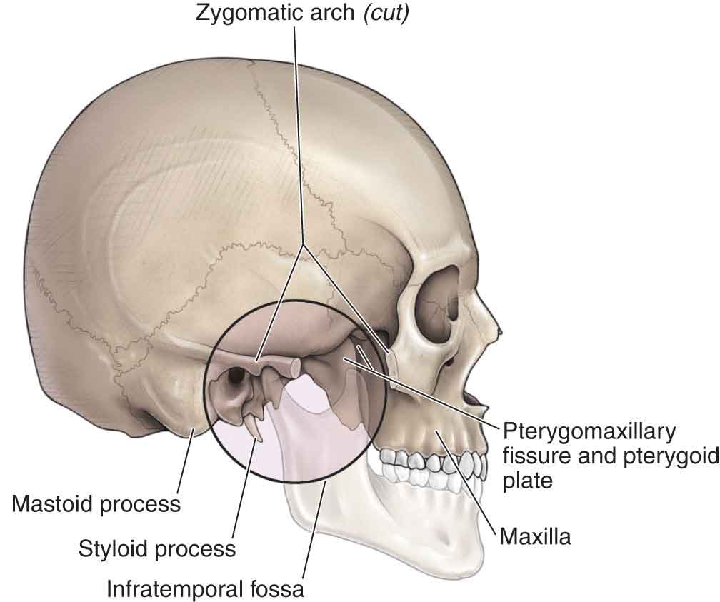
 Normal Anatomy Of The Jaw This Lateral View Of The Skull
 Temporomandibular Joint Wikipedia
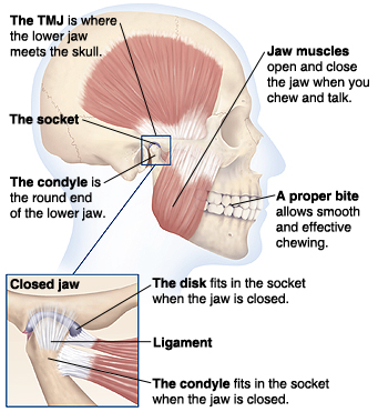
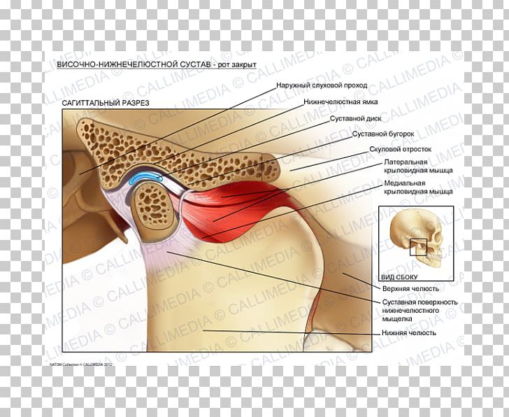
 Surface Anatomy Of Temporomandibular Joint Mouth Closed
 Diagnosis And Treatment Of Temporomandibular Disorders
 Nras National Rheumatoid Arthritis Society
 Temporomandibular Tmj Joint And Its Disorder Osteopathy
 Tmj Massage Treatments For Jaw Headache Pain Book Online Now

 Temporomandibular Joint Tmj Disorders An Overview
 Tmj Leawood Cosmetic Family Dentistry Pediatric
 Temporomandibular Joint Tmj Anatomy Animation
 Tmj Jaw Pain T O P S Physical Therapy
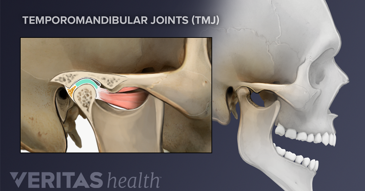
 Temporomandibular Joint Dysfunction Anatomy Lateral
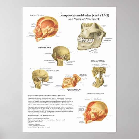 Tmj Anatomy Muscular Attachments Chart Doctor
![]() The Temporomandibular Joint Tmj Anatomy And Supply Kenhub
The Temporomandibular Joint Tmj Anatomy And Supply Kenhub
Belum ada Komentar untuk "Anatomy Of Temporomandibular Joint"
Posting Komentar