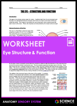
Anatomy Of Eye And Functions
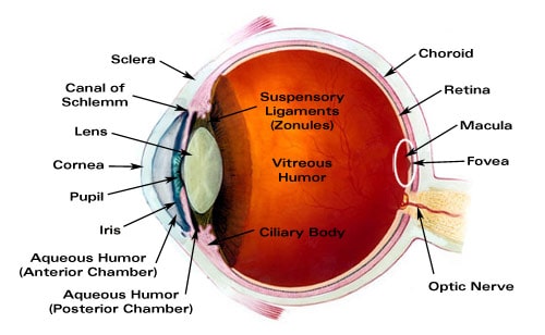
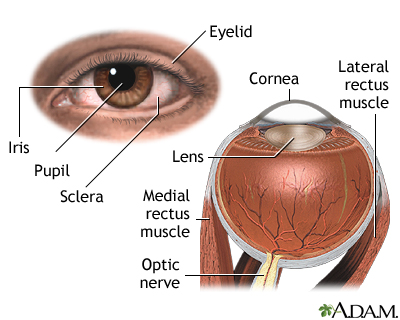
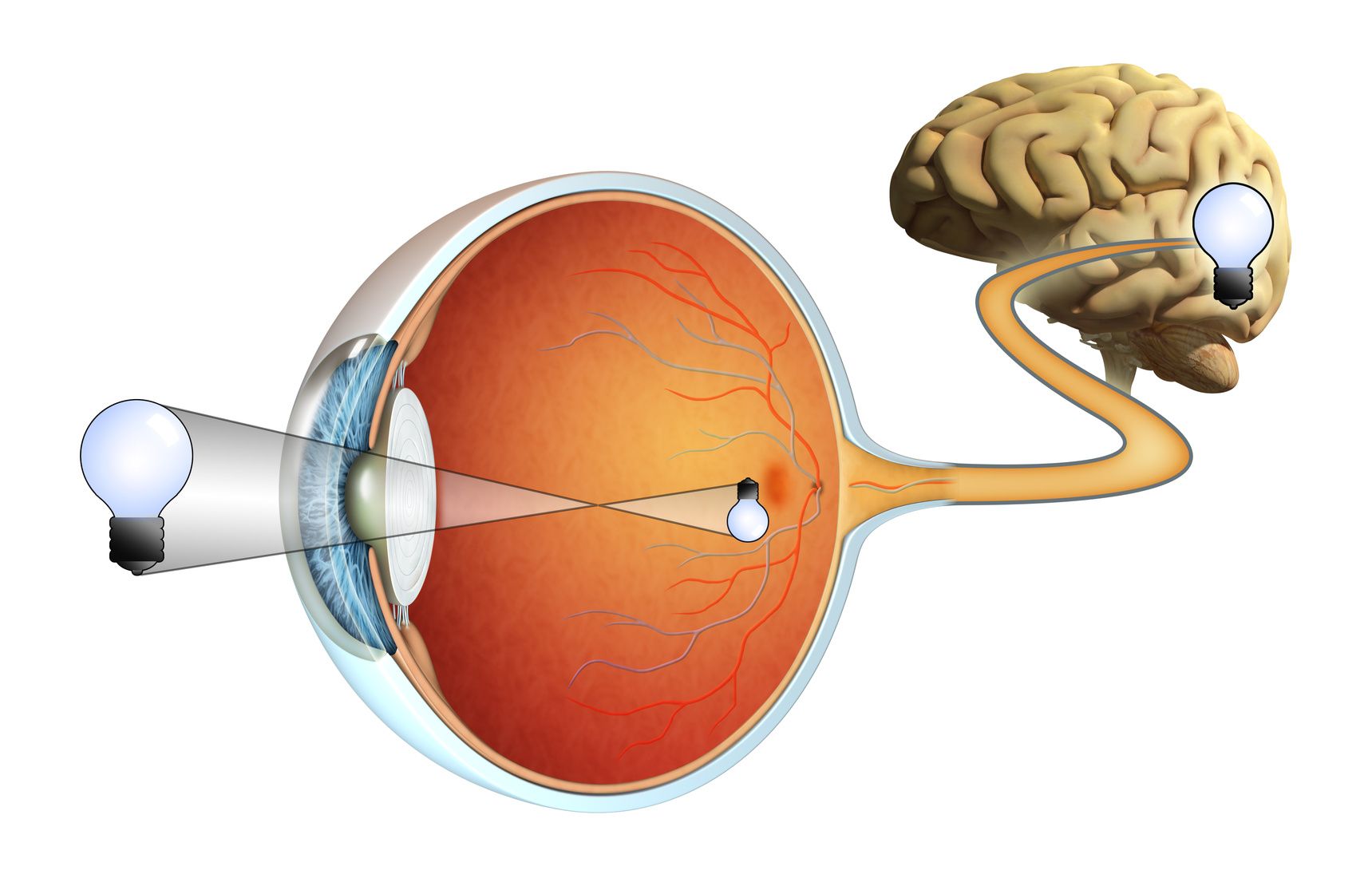
The outer layer of the eyeball is a tough white opaque membrane called the sclera the white of the eye. Light enters through the cornea past the iris through the pupil refracted by the lens and onto the retina of the eye.
 The Anatomy Of The Eye Anterior Segment Precision Family
The slight bulge in the sclera at the front of the eye is a clear thin dome shaped tissue called the cornea.
Anatomy of eye and functions. The white part of the eye that one sees when looking at oneself in the mirror is. Light path through the eye. Anatomy and physiology of the eye conjunctiva.
The nerve at the back of the eye that transports electric signals to the brain. Cornea the clear front window of the eye which transmits and focuses ie. The middle layer is the choroid.
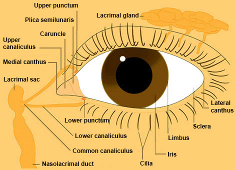
The iris or coloured part of the eye surrounds the pupil. The pupil or black dot at the centre of the eye is an opening through which light can enter. This dome shaped layer protects your eye from elements that could cause damage to the inner parts of the eye.
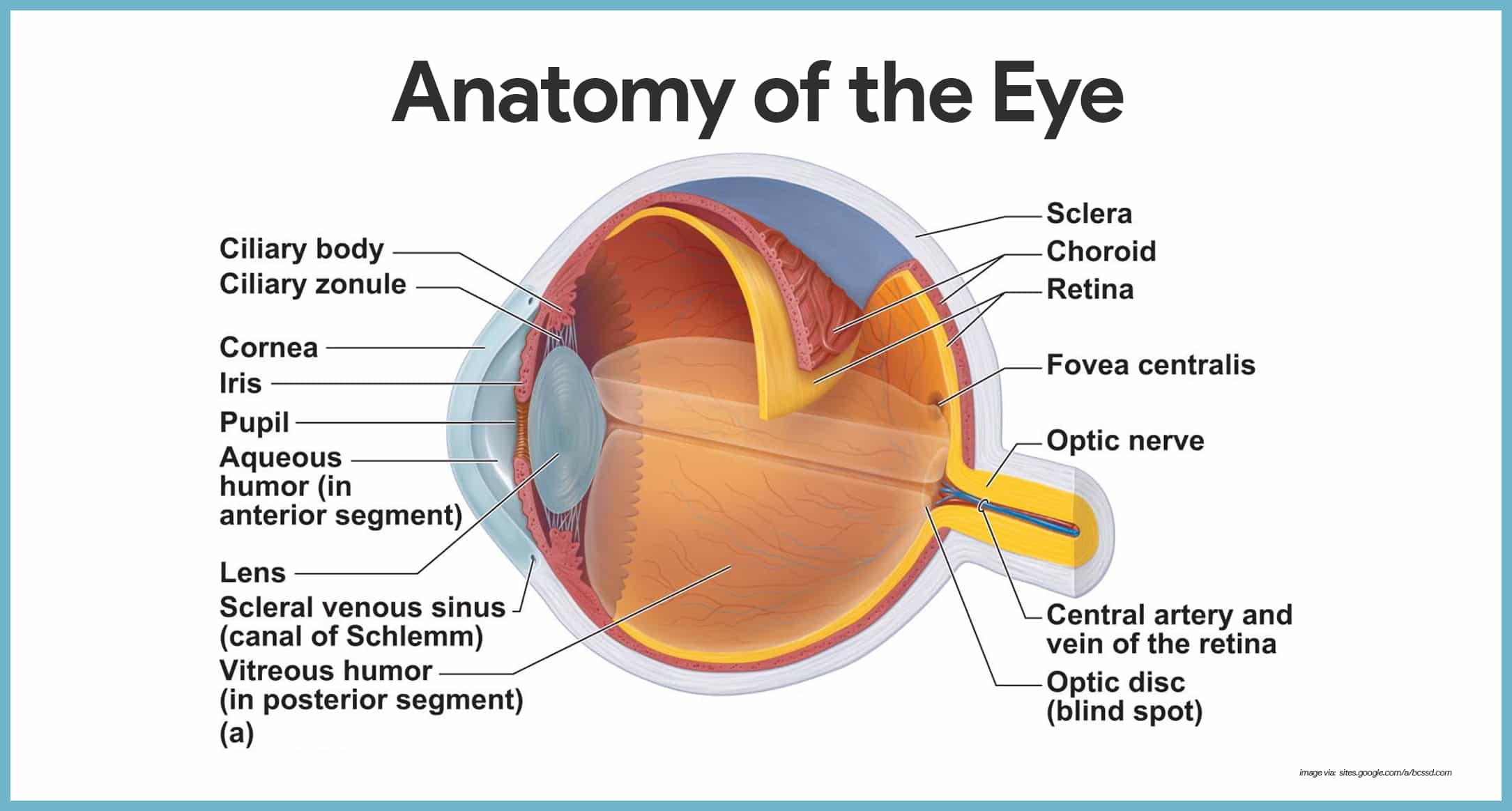
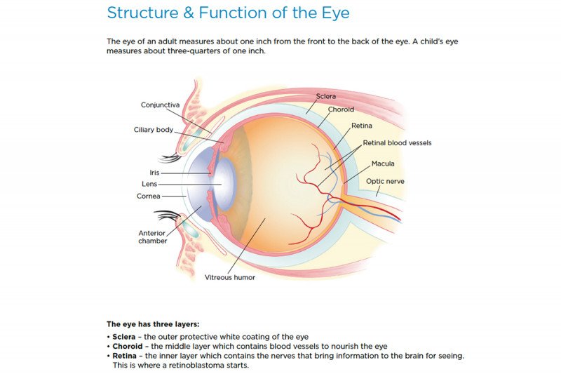
Anatomy of the eye choroid layer containing blood vessels that lines the back of the eye and is located between. The macula is a small extra sensitive area in the retina that gives you central vision. There are several layers of the cornea creating a tough layer that provides additional protection.
Eye color is created by the amount and type of pigment in your iris. Cornea the cornea is the outer covering of the eye. Eye parts and functions.
The conjunctiva is a thin transparent layer of tissues covering the front of the eye. The transparent orb within the eye that refracts light to the retina of the eye. Ciliary body structure containing muscle and is located behind the iris which focuses the lens.
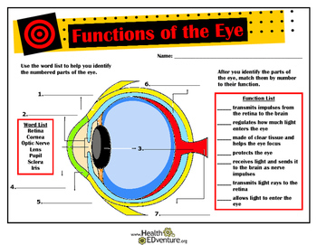
The eye has many parts that must work together to produce clear vision. The iris of the eye functions like the diaphragm of a camera controlling the amount of light reaching the back of the eye by automatically adjusting the size of the pupil aperture. Multiple genes inherited from each parent determine a persons eye color.
The surface of the eye and of the inner eyelids is covered by a clear protective membrane called the conjunctiva. The cornea is the transparent clear layer at the front and. Light is focused primarily by the cornea the clear front surface of the eye which acts like a camera lens.
The front of the choroid is the colored part of the eye called the iris. Parts of the eye and their functions. The eye has many parts which work together to accomplish vision and to keep the structures required for vision safe from infection and injury.
Behind the eye your optic nerve carries these impulses to the brain. The sclera or white part of the eye protects the eyeball.
 Major Ocular Structures Laramy K Independent Optical Lab
 Special Senses Anatomy And Physiology Nurseslabs
 Parts Of The Eye And Their Functions Video Lesson
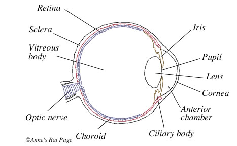 The Rat S Eyes
 Vision Anatomy And Physiology
 Eye Anatomy Glaucoma Research Foundation
 Neuroscience For Kids The Eye And Its Connections
 Vision Anatomy And Physiology
Eye Anatomy Structure Function Of Vision Hs Ls1 A By
 Human Eye Wikipedia
/GettyImages-695204442-b9320f82932c49bcac765167b95f4af6.jpg) Structure And Function Of The Human Eye
Structure And Function Of The Human Eye
 Human Eye Anatomy And Its Function
 Human Eye Anatomy Structure And Function
 External And Internal Eye Anatomy Medlineplus Medical
 Blind Spot Anatomy Britannica
 Frontiers Frontal Eye Field Where Art Thou Anatomy
 Functions And Anatomy Of The Eye
 Cataracts Cleveland Clinic
 Eye Structure And Function In Dogs Dog Owners Merck
 Anatomy And Function Of The Eye
 Anatomy Of The Eye Children S Wisconsin
 Eye Anatomy And Function
 Anatomy And Structure Of The Eye Brightfocus Foundation
 Eye From Front Anatomy The Eyes Have It
 Eye Structure And Function In Dogs Dog Owners Merck
 Eye Anatomy And Vision Course Hero
 Anatomy Of The Eye The Ottawa Hospital
 Retinoblastoma Anatomy Of The Eye Memorial Sloan


Belum ada Komentar untuk "Anatomy Of Eye And Functions"
Posting Komentar