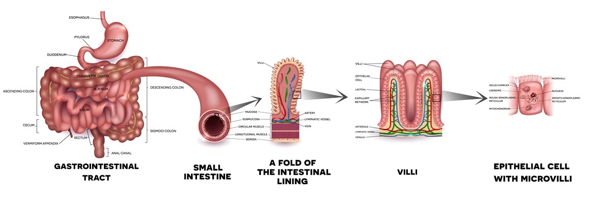
Small Bowel Anatomy
After death this length can increase by up to half. It extends from the pylorus of the stomach to the ileocaecal junction where it meets the large intestine at the ileocaecal valve.
 Imaging Anatomy Of Small Intestine
The ileum joins the cecum the first portion of the large intestine at the ileocecal sphincter or valve.

Small bowel anatomy. The small intestine latin. The duodenum jejunum and ileum. It is about 67 to 76 metres 22 to 25 feet long highly convoluted and contained in the central and lower abdominal cavity.
It lies between the stomach and large intestine and receives bile and pancreatic juice through the pancreatic duct to aid in digestion. Anatomy structure and pathology of the small intestine small bowel see online here. The small intestine is a highly coiled tubular structure that forms the end site of digestion.
The small intestine or small bowel is an organ in the gastrointestinal tract where most of the end absorption of nutrients and minerals from food takes place. The small intestine small bowel is about 20 feet long and about an inch in diameter. Together with the esophagus large intestine and the stomach it forms the gastrointestinal tract.
Intestinum tenue spans a range of about 35 m from the pylorus of the stomach to the bauhins valve located at the passage to the colon. It is thicker more vascular and has more developed mucosal folds than the jejunum. It has distinctive mucosal folds valvulae conniventes and is made up of three functional units.
The intestines include the small intestine large intestine and rectum. Anatomically the small bowel can be divided into three parts. It is the region where most digestion and absorption of food takes place.
It is the most important part of the alimentary canal and leads to the large intestine. It is approximately 65m in the average person and assists in the digestion and absorption of ingested food. Aprof frank gaillard et al.
In living humans the small intestine alone measures about 6 to 7 meters long. Sometimes this organ is also called small bowel. Small intestine a long narrow folded or coiled tube extending from the stomach to the large intestine.
It has a surface area of over 200 meters. The small intestine is made up of the duodenum jejunum and ileum. The small bowel or small intestine is the section of bowel between the stomach and the colon.
The ileum is the longest part of the small intestine measuring about 18 meters 6 feet in length. The small intestine is an organ located within the gastrointestinal tract. The first and middle sections of the small intestine the small intestine is the section of your digestive tract where the majority of food digestion and nutrient absorption takes place.
 Small Bowel Resection Series Normal Anatomy Medlineplus
 Small Intestine Anatomy Britannica
 The Small Intestine Duodenum Jejunum Ileum
 23 5 The Small And Large Intestines Anatomy And Physiology
 Small Bowel Prolapse Enterocele Disease Reference Guide
 Anatomy Small Intestine Small Bowel Ppt Download
 Anatomy Of The Small Intestine
 Bowel Obstruction Ucsf Fetal Treatment Center
 Difference Between Small Intestine And Large Intestine With
 Intestinal Obstruction Symptoms And Causes Mayo Clinic
 Small Bowel Obstruction Cleveland Clinic
 What Does The Small Intestine Do
 Imaging Anatomy Of Small Intestine
 Large And Small Intestine Anatomy
 Celiac Or Coeliac Disease As An Intestine Anatomy Medical
 Small Intestine Anatomy Location And Function Kenhub
 23 5 The Small And Large Intestines Anatomy And Physiology
 Small Intestine Anatomy Digital Illustration Stock Photo
 Inflammatory Bowel Disease Ibd Symptoms And Causes
 Small Intestine Anatomy Overview Gross Anatomy
 Small Intestine Drawing At Getdrawings Com Free For
 Radiological Anatomy Small Intestine Stepwards
 Gross And Microscopic Anatomy Of The Small Intestine
 Regions Of The Small Intestine The Duodenum Is Attached To
 Small And Large Intestine Johns Hopkins Division Of


Belum ada Komentar untuk "Small Bowel Anatomy"
Posting Komentar