Infant Skull Anatomy
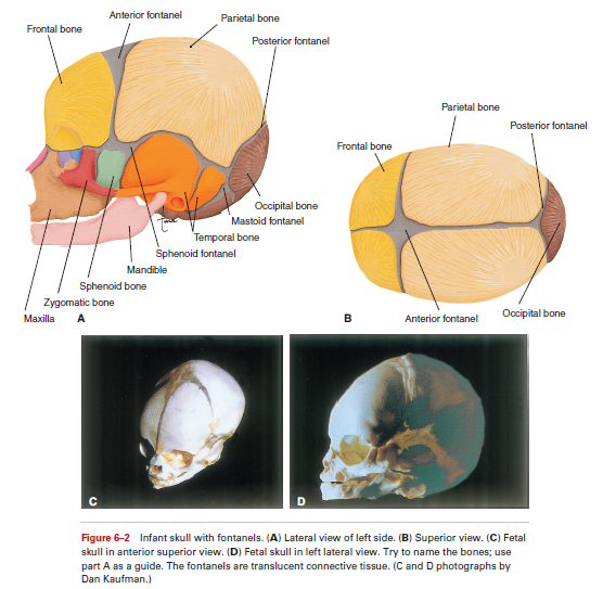
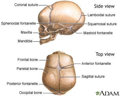
The infant skulls bones are separated by fontanelles or soft spots. In an infant the skull bones are unossified occipital temporal sphenoid frontal mandible bones have more than one piece.
 Human Anatomy Life Size Adult And Baby Skull Models Adult
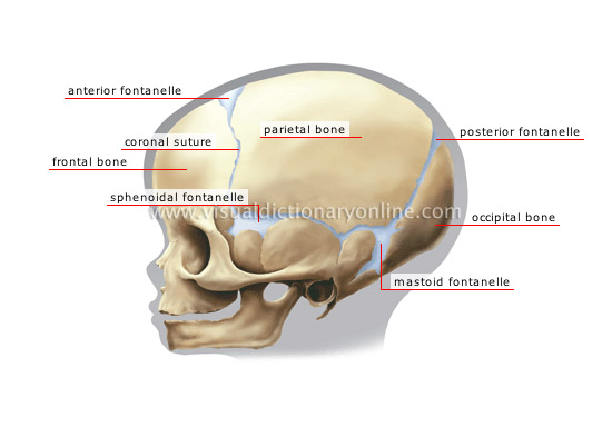
Fetal skull anatomy is also characterized by the presence of soft membranous areas called fontanelles soft spots that eventually become sutures in an adult skull.
Infant skull anatomy. The skull develops as a loosely joined system of bones formed in the soft tissue matrix surrounding the brain. Infant and child skulls are considerably pliable due to the segmental development and arrangement of the skull bones plus the flexibility of individual bones which are extremely thin. These spaces are called fontanels or soft spots.
These bony plates cover the brain and are held together by fibrous material called sutures. Anatomy of the newborn skull. Although the skull appears to be 1 large bone there are actually several major bones that are connected together.
At the brain case these elements are separat. The major bones that compose the skull of a newborn include the following. Although the skull appears to be 1 large bone there are actually several major bones that are connected together.
Anatomy of the newborn skull. During the first few years of life these bones are not fused but held together by a type of stretchy tissue called cranial sutures. At birth the skull is composed of 44 bony elements that eventually fuse and increase in hardness.
These membranous areas are called fontanels. The major bones that compose the skull of a newborn include the following. Although the skull appears to be one large bone there are actually several major bones that are connected together.
The major bones that compose the skull of a newborn include the following. They permit some movement between the bones so that the developing skull is partially compressible and can slightly change shape. The entire infant skull is composed of numerous pieces that are essentially softer than the adult skull.
At birth the skull is incompletely developed and fibrous membranes separate the cranial bones. There are two spaces in the skull that are not covered by bone but only by the cranial sutures. Anatomy of the newborn skull.
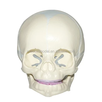
An infants skull is made up of six bones.
 How Much Do Babies Skulls Get Squished During Birth A
 Solved What Is The Difference Between The Frontal Bone Of
 A Further Look At Head Anatomy Kerry Mccormick Animation

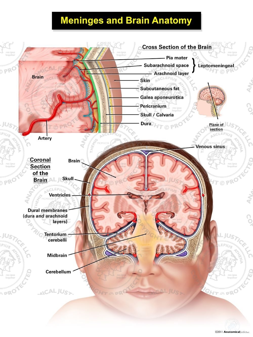
Meninges Of The Brain Infant
The Skull Anatomy And Physiology Openstax
 Details About Double Head Baby Skull Human Research Model Infant Skeleton Anatomical Brain
 The Skull And Foramina Human Anatomy
 The Skull Boundless Anatomy And Physiology
 Infant Skull Facial Bones Axial Skeleton Body Anatomy
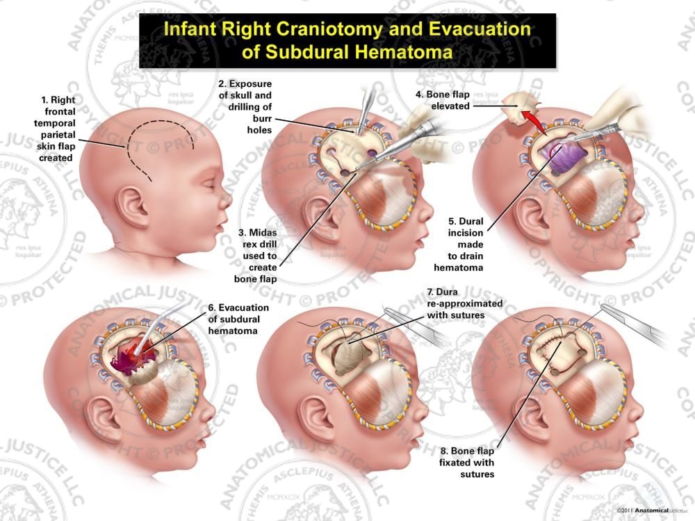 Infant Right Craniotomy And Evacuation Of Subdural Hematoma
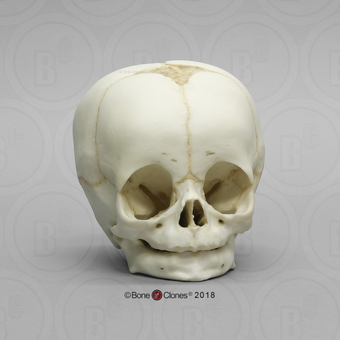 4 Month Old Human Child Skull Bone Clones Inc
 Skull Of A Newborn Medlineplus Medical Encyclopedia Image
The Skull Anatomy And Physiology Openstax
 Foetal Child Skull Infant Skull Model
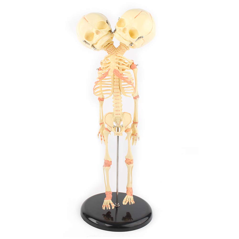 Beautylady Double Head Baby Skull Model Double Headed Infant
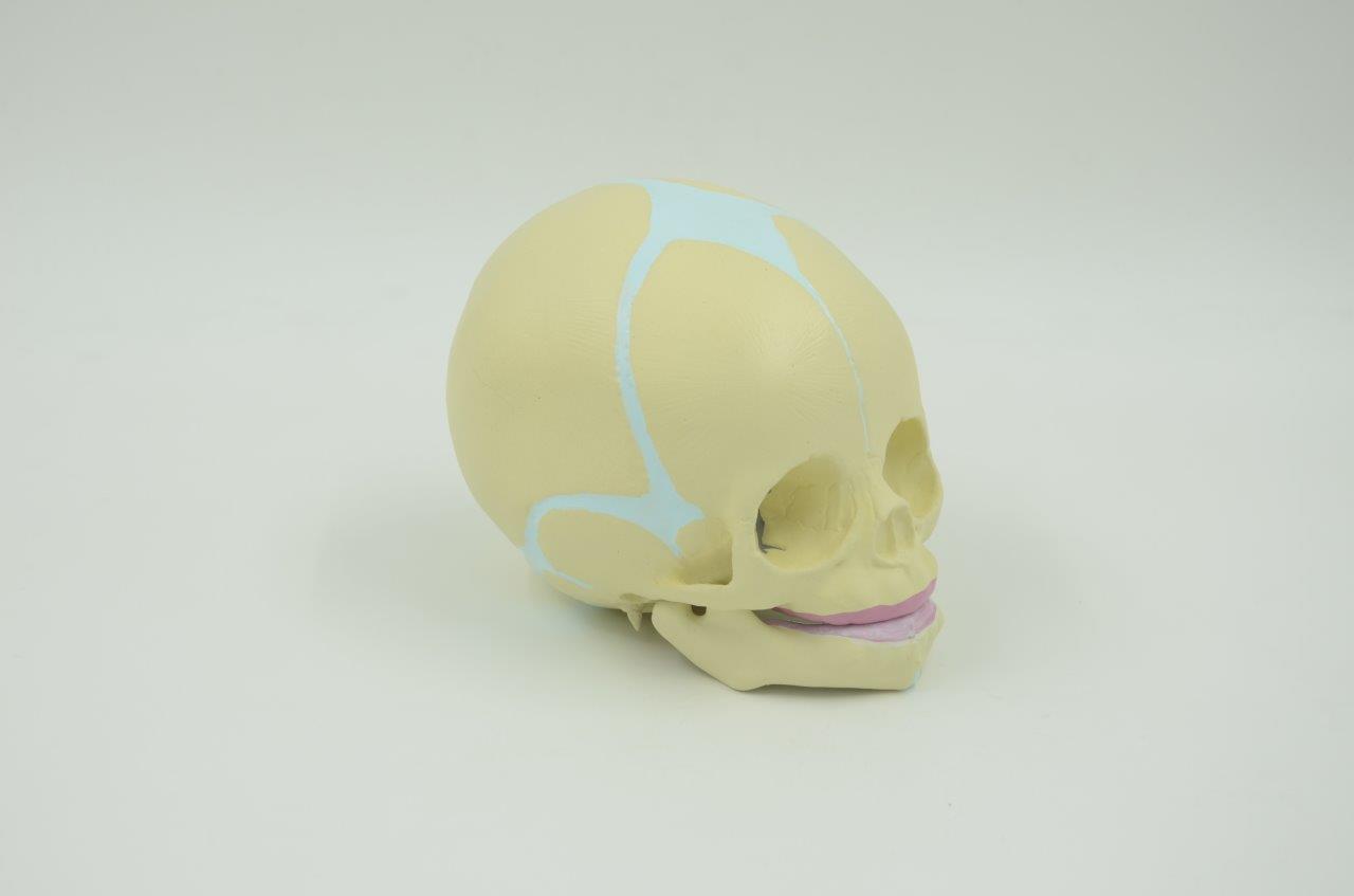 Baby Skull Model Fetus Skull
 Baby Skull Lateral View In 2019 Skull Anatomy Skeleton
 Skeletal System Anatomy And Physiology Nurseslabs
 Human Fetal Baby Infant Medical Anatomical Skull Model For
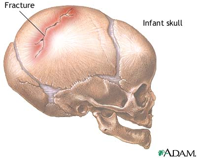 Infant Skull Fracture Medlineplus Medical Encyclopedia Image
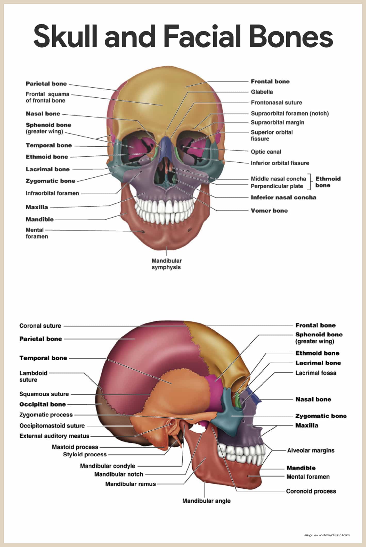
 Skull Cranium And Facial Bones
 Infant Skull Model Baby Skull Model Buy Infant Skull Baby Skull Baby Skull Model Product On Alibaba Com
 Double Headed Three Eyes Infant Skeletal Model Skull Skeleton Anatomical Brain Anatomy Education Model
 Artificial Infant Skull Model
Belum ada Komentar untuk "Infant Skull Anatomy"
Posting Komentar