Eye Anatomy Iris
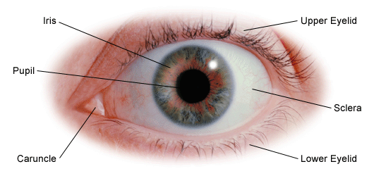
The iris is a flat and ring shaped membrane behind the cornea of the eye with an adjustable circular opening in the center called a pupil. Muscles in the iris dilate widen or constrict narrow the pupil to control the amount of light reaching the back of the eye.
 Eye Anatomy Eyes On J Optometry
Multiple genes inherited from each parent determine a persons eye color.
Eye anatomy iris. Anatomy of the eye. Parts of the eye. And when there is low light the iris opens up the pupil to let in more light.
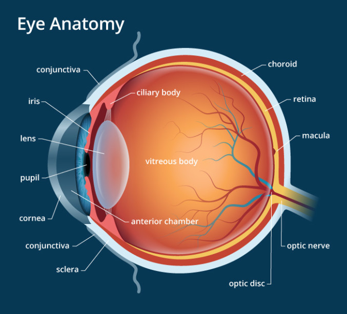
The anatomy of the eye is fascinating and this quiz game will help you memorize the 12 parts of the eye with ease. Light enters our eyes through the pupil then passes through a lens and the fluid filled vitreous body before it is projected onto the retina. The macula is a small extra sensitive area in the retina that gives you central vision.
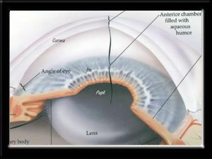
Iris the colored part of the eye which helps regulate the amount of light entering the eye. Lens focuses light rays onto the retina. It is bathed in front and behind by a fluid known as the aqueous humour.
This is the structure that provides an individual with eye color. Behind the anterior chamber is the eyes iris the colored part of the eye and the dark hole in the middle called the pupil. Iris in anatomy the pigmented muscular curtain near the front of the eye between the cornea and the lens that is perforated by an opening called the pupil.
In optical terms the pupil is the eyes aperture while the iris is the diaphragm. Behind the eye your optic nerve carries these impulses to the brain. The eyes crystalline lens is located directly behind the pupil and further focuses light.
Eye color is created by the amount and type of pigment in your iris. In humans and most mammals and birds the iris is a thin circular structure in the eye responsible for controlling the diameter and size of the pupil and thus the amount of light reaching the retina. The iris is located in front of the lens and ciliary body and behind the cornea.
Eye color is defined by that of the iris. Although the eye is small only about 1 inch in diameter each part plays an important role in allowing people to see the world. When there is bright light the iris closes the pupil to let in less light.
Together with the pupil the iris is responsible for regulating the amount of light that gets into the eye. The iris of the eye functions like the diaphragm of a camera controlling the amount of light reaching the back of the eye by automatically adjusting the size of the pupil aperture. Anatomy of the eye.
The eye is surrounded by the orbital bones and is cushioned by pads of fat within the orbital socket.
 Wikipremed

 Eye Anatomy A Closer Look At The Parts Of The Eye

Parts Of The Eye American Academy Of Ophthalmology
 How The Eye Works Fighting Blindness
 Human Eye Iris Vector 3d Realistic Eyeball Illustration
 Eye Anatomy Iris Optic Pupil Vision Stock Vector Royalty
 Anatomy Of The Eye The Ottawa Hospital
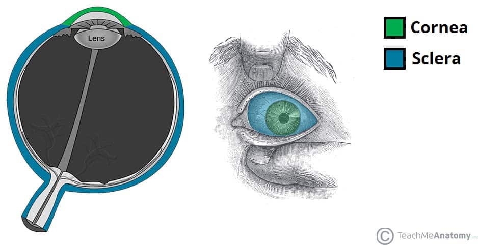
The Eyeball Structure Vasculature Teachmeanatomy
 Human Eye Anatomy Structure Medical Infographics For Ophthalmology
 The Iris Its Anatomy Function Related Eye Diseases

 Anatomy Of The Eye 101 Eyecheck
 Educational Pillow Sham Human Eye Anatomy Cornea Iris Pupils
 Anatomy Of The Eye
 Eye Anatomy Pullen Eye Care
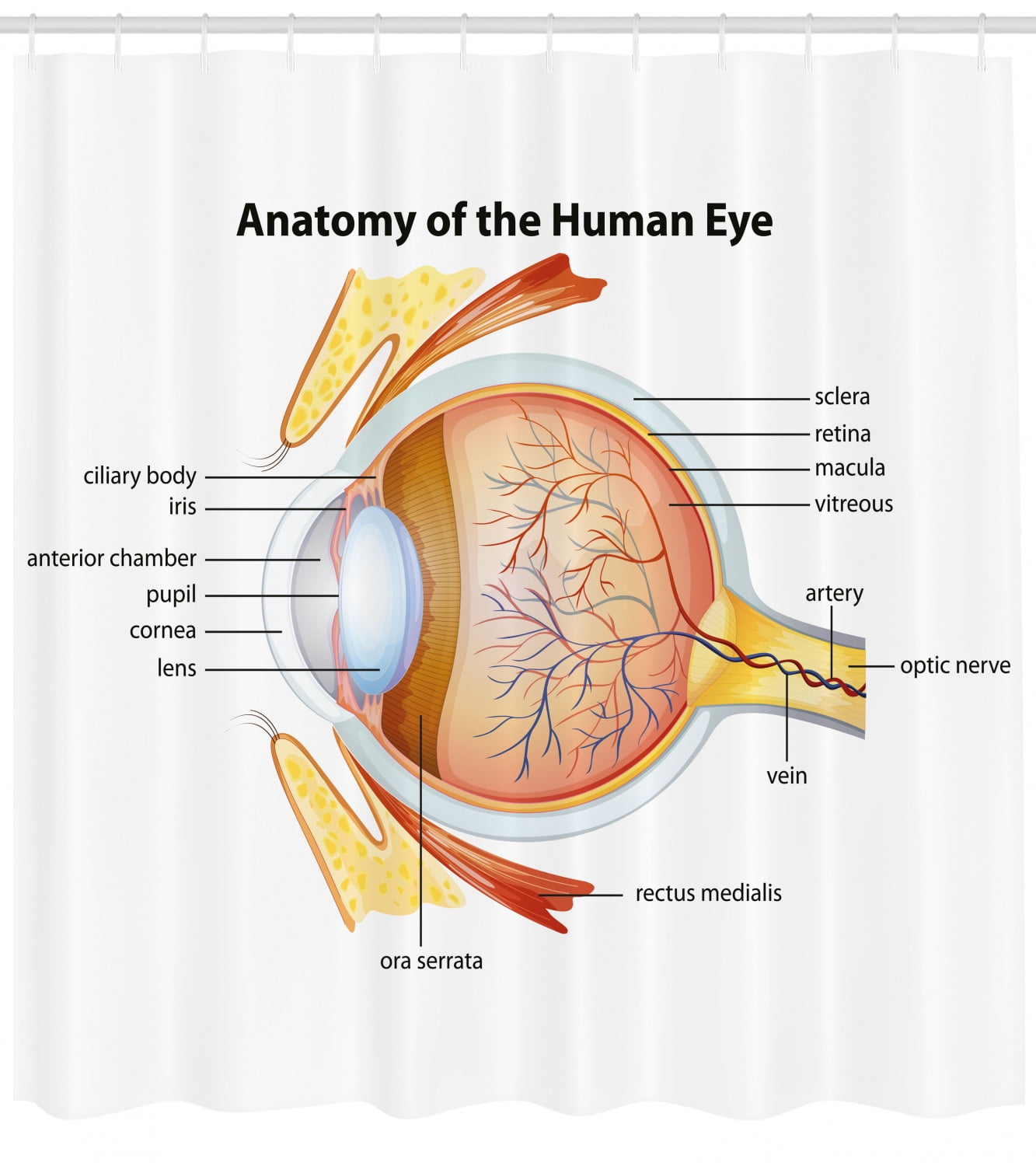 Educational Shower Curtain Human Eye Anatomy Cornea Iris Pupils Optic Nerves Graphic Print Fabric Bathroom Set With Hooks 69w X 75l Inches Long

:max_bytes(150000):strip_icc()/GettyImages-695204442-b9320f82932c49bcac765167b95f4af6.jpg) Structure And Function Of The Human Eye
Structure And Function Of The Human Eye
 Eye Anatomy Ozarks Family Vision Centre
 Amazon Com Eye Anatomy Watercolor Poster Art Print Human
 Iris And Uvea Of The Eye Allaboutvision Com
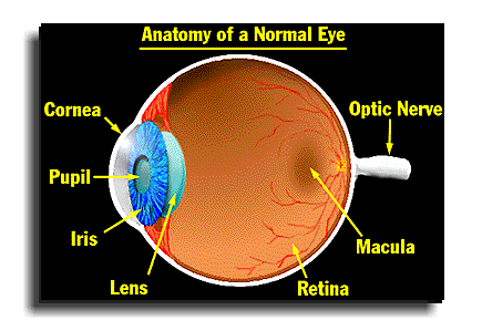 Anatomy Of A Normal Human Eye Amdf
Belum ada Komentar untuk "Eye Anatomy Iris"
Posting Komentar