Anatomy Of The Aortic Valve
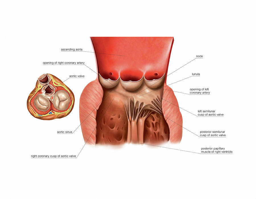
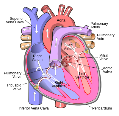
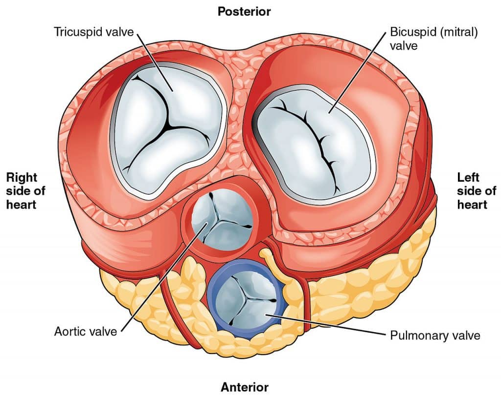
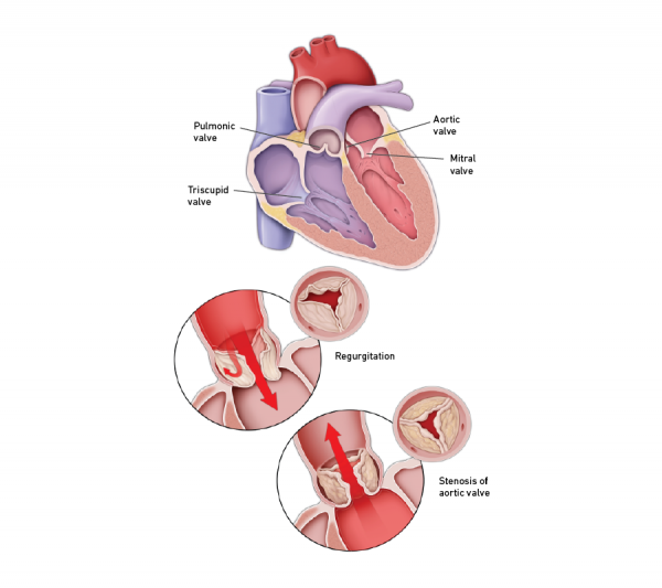
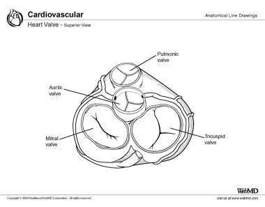
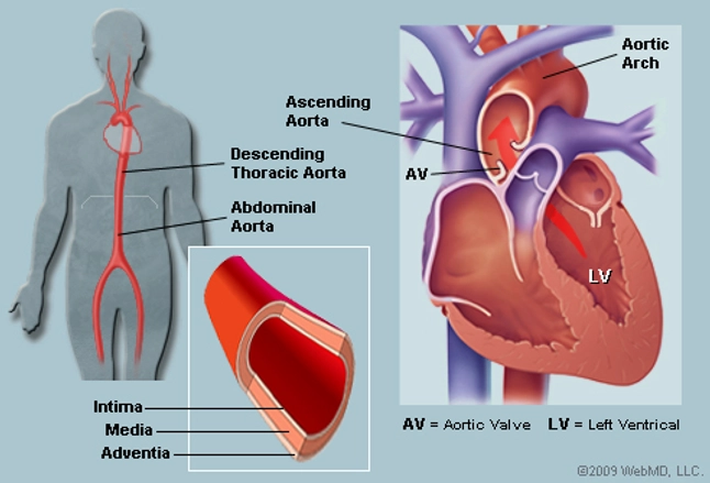
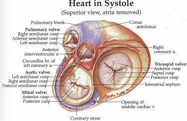
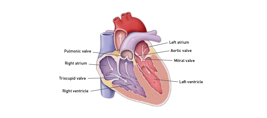
The aortic valve is a part of the aortic root complex which is composed of the sinuses of valsalva the fibrous interleaflet triangles and the valvular leaflets themselves. The relative positions of the aortic mitral pulmonary and tricuspid valves are shown in the diagram of the heart at the center of the figure.
 Chapter 31 Pathophysiology Of Aortic Valve Disease
Anatomy of the aortic valve.
:max_bytes(150000):strip_icc()/human-heart-circulatory-system-598167278-5c48d4d2c9e77c0001a577d4.jpg)
Anatomy of the aortic valve. With each of the heart muscles contractions oxygenated blood exits the left ventricle through the aortic valve. The lamina fibrosa is the widest layer and faces the aortic or arterial side of the valve cusp. The aortic valve cusps have 3 readily identifiable layers.
The aortic valve is located between the aorta and the hearts left ventricle. The aortic valve is composed of 4 key parts. It functions as a one way valve.
The pulmonary vein delivers oxygenated blood to the hearts left atrium. The left coronary cusp lcc the right coronary cusp rcc and the non coronary cusp ncc. The aortic valve has three cusps.
When the heart relaxes between contractions the aortic valve closes preventing blood in the aorta from returning to the left ventricle. The aorta is the largest artery in the body. Approximately two thirds of the circumference of the lower part of the aortic root is connected to the septum and the remaining third is connected via a fibrous continuity known as the aortic mitral curtain to the mitral valve.
1 lamina fibrosa 2 lamina spongiosa and 3 lamina radialis. Posteromedial trigone right carpentiers reconstructive valve surgery ch 5 2010. Anatomical terminology edit on wikidata the aortic valve is a valve in the human heart between the left ventricle and the aorta.
Then it passes through the mitral valve and into the left ventricle. It is one of the two semilunar valves of the heart the other being the pulmonary valve. The valve comprises of the semilunar leaflets with attachments or hingelines to ventricular and aortic walls and the anterior mitral leaflet interleaflet triangles aortic sinuses of valsalva and the sinutubular junction.
The heart pumps blood from the left ventricle into the aorta through the aortic valve. Three leaflets on the aortic valve open and close with each heartbeat to allow one way flow of blood. Ventricular systole is a very short amount of time during the cardiac cycle for the leaflets to open and close allowing blood to exit out of the heart.
When the left ventricle contracts the valve only allows blood to flow out of the left ventricle and into the aorta. Surgical anatomy aortic root is anchored between the pulmonary root anteriorly and mitral and tricuspid posteriorly fibrous skeleton of the heart pulmonary. The aorta begins at the top of the left ventricle the hearts muscular pumping chamber.
The aortic valves main function is to act as a gateway for blood to exit out of the ventricle during systole and push through the aorta for the body to receive oxygenated blood. To appreciate the anatomy of the aortic valve it is necessary to examine its component parts and how they relate to each other so as to function as a unit.
 Aortic Valve Of The Heart
 Standardized Imaging For Aortic Annular Sizing Implications
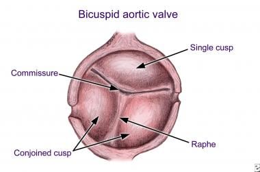
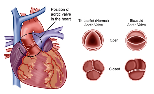 Bicuspid Aortic Valve Disease Cardiac Health
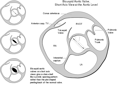 Bicuspid Aortic Valve Wikipedia
 Aortic Valve Anatomy Implications For Transcatheter Aortic

Av And Semilunar Heart Valves
 Figure 4 From Gross Anatomy Of The Aortic Valve Of
 Aortic Valve Wikipedia
 The Anatomy Of Aortic Valve Aortic Root Coronary Arteries
 Aortic Valve Anatomy Implications For Transcatheter Aortic
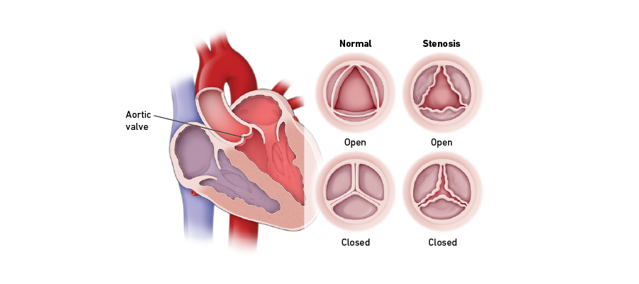 Aortic Valve Stenosis Temple Health
 Whether You Need Surgery To Replace Your Aortic Valve Due To
 The Heart Valves Tricuspid Aortic Mitral Pulmonary
 Anatomy Of The Aortic Valve Longitudinal Section Lvot

 Heart Valve Diseases Temple Health
 Imaging Of The Aortic Valve With Mri And Ct Angiography
 Bicuspid Aortic Valve Background Pathophysiology Epidemiology
 Aortic Valve Anatomy Overview Gross Anatomy Microscopic

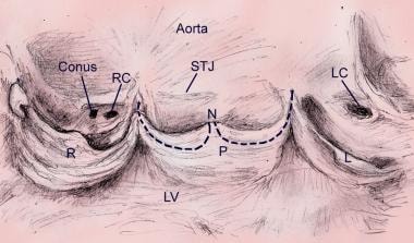
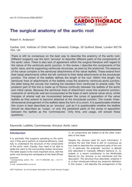 The Surgical Anatomy Of The Aortic Root Multimedia Manual
 Aortic Stenosis Causes Symptoms And Progression What Is
 Aortic Valve Anatomy Overview Gross Anatomy Microscopic
 Bicuspid Aortic Valve Disease Savr Or Tavr
 The Aorta Human Anatomy Picture Function Location And
 Heart Model With Valves
 Aortic Valve Stenosis Temple Health
 Aortic Valve Anatomy The Aortic And Pulmonary Valves Are
Chapter 1 Surgical Anatomy Of The Aortic And Mitral Valves
Belum ada Komentar untuk "Anatomy Of The Aortic Valve"
Posting Komentar