Foot Anatomy Veins
It is accompanied by the dorsalis pedis vein. The veins of the foot circulate oxygen depleted blood from the tissues back to the heart.
 Dorsal Digital Veins Of The Foot Wikipedia
Anatomy of the foot perforator veins.
Foot anatomy veins. The small saphenous great saphenous and anterior tibial veins. Pain swelling redness and bruising may be signs of a fracture. Gait at the beginning of a step the distal calf pump is activated.
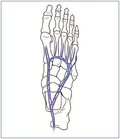
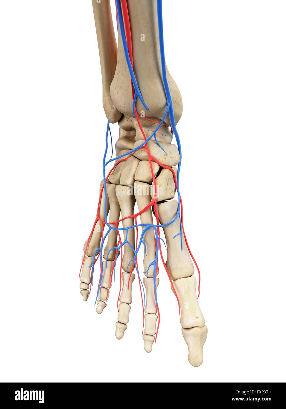
These foot perforator veins are split into two well separated functional units medial and lateral connected to each plantar vein. A viral infection in the sole of the foot that can form a callus with a central dark spot. Deoxygenated blood returning from the tissues of the feet is collected by many veins that join to form the dorsal venous arch on the top of the foot and the deep plantar venous arch of the sole of the foot.
Foot perforator veins provide direct connections between the plantar veins and the roots for both saphenous systems. There are medial and lateral marginal veins which drain both of the dorsal and plantar parts of the specific sides within the dorsal venous arch alongside the foot. The anterior compartment muscles contract dorsiflect the foot and empty its veins ie the anterior tibial veins.
Blood from the dorsal venous arch passes into three major veins in the leg. One common problem is varicose veins. It interacts along with proximally situated dorsal venous network and receives the dorsal digital as well as dorsal metatarsal veins.
The metatarsal bones are the most frequently broken bones in the feet either from injury or repetitive use. Circulation problems of the foot are common in both the elderly and obese people as well as those who stand for long periods of time. Venous foot pump voiding.
This process is initiated by dorsiflexion of the foot as the leg is lifted to take a step.
![]() Veins Of The Lower Limb Anatomy Kenhub
Veins Of The Lower Limb Anatomy Kenhub
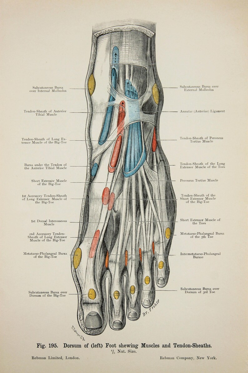
 Ankle Foot Anatomy
 Schematic View Of Venous Anatomy From Insightful Phlebology
 Amazon Com Anatomy Ankle Vein Knee Print Sra3 12x18
 Great Saphenous Vein Anatomy Pictures And Information
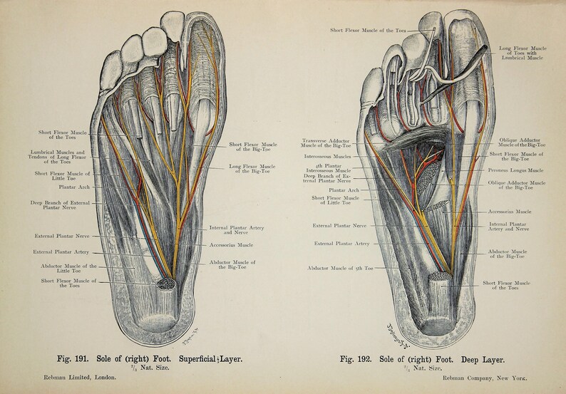
 1831 Foot Anatomy Foot Muscles Nerves Illustration Muscles Veins Arteries Print Human Body Poster Bourgery Medicine Wall Art
 Phalanges Approach Dorsal Approach To P1 Phalanges

 Amazon Com Anatomy Foot Vein Tendon Print Sra3 12x18
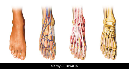
 Human Foot Anatomy Cutaway Representation Showing Skin Veins
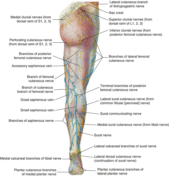
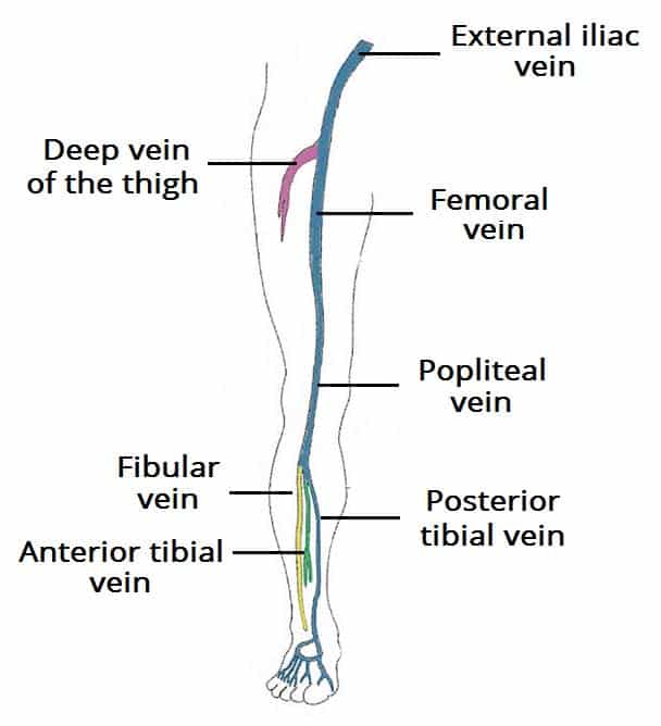 Venous Drainage Of The Lower Limb Teachmeanatomy
 The Venous System Of The Foot Anatomy Physiology And
 Ankle Foot Anatomy
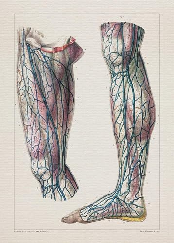
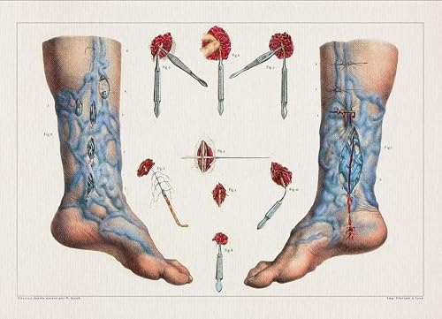
 Feet Ankles Tendons Arteries Veins Nerves C 1900 Double Sided Antique Anatomy Print Colour Anatomical Print Lithograph
 Amazon Com Anatomy Varicose Veins Foot Print Sra3 12x18
 Spinal Veins An Overview Sciencedirect Topics


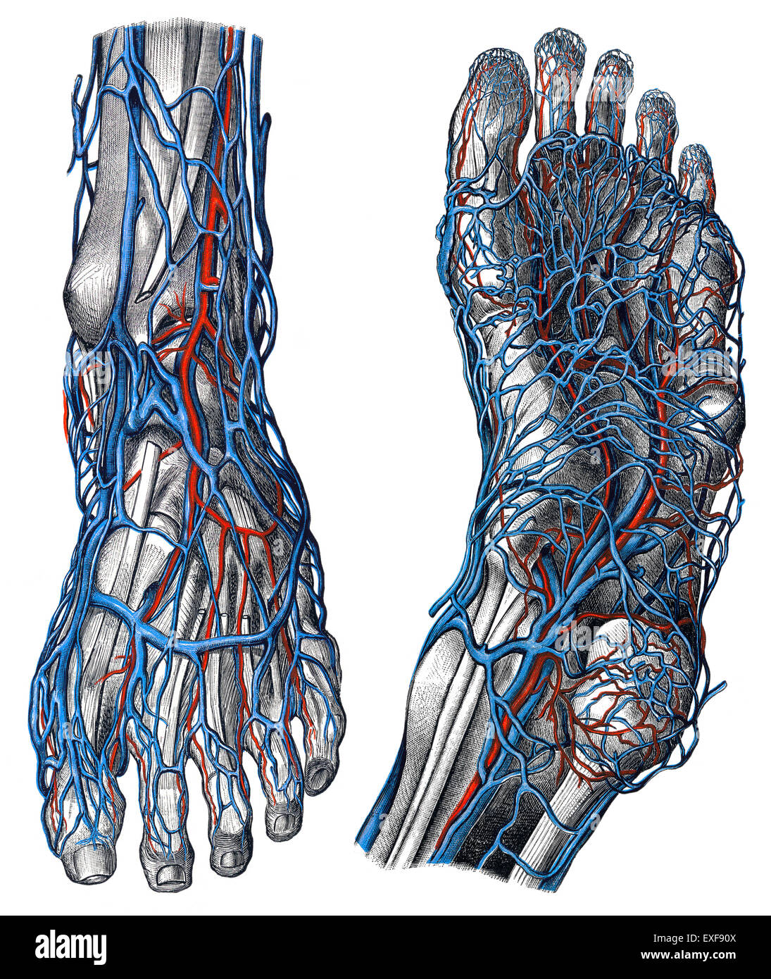 The Veins Of The Foot Stock Photo 85158890 Alamy

![]() Small Saphenous Vein Anatomy Kenhub
Small Saphenous Vein Anatomy Kenhub
 Image Result For Posterior Leg Muscles Muscle Foot
 Feet Ankles Tendons Arteries Veins Nerves C 1900 Double Sided Antique Anatomy Print Colour Anatomical Print Lithograph
 Anatomy Springerlink
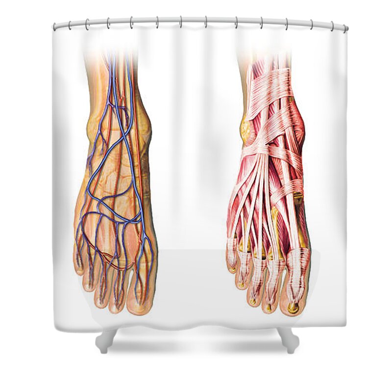 Human Foot Anatomy Showing Skin Veins Shower Curtain
 Veins Arteries Foot Stock Photos Veins Arteries Foot Stock

 Human Foot Anatomy Cutaway Representation Showing Skin
 Human Foot Anatomy Showing Skin Veins Metal Print
Belum ada Komentar untuk "Foot Anatomy Veins"
Posting Komentar