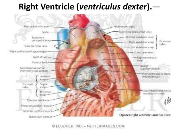
Anatomy Of Right Ventricle
It also marks the inferior border of the cardiac silhouette. The right ventricle is one of the hearts four chambers.
 Heart And Right Ventricle
The normal rv is anatomically and functionally different from the left ventricle which precludes direct extrapolation of our knowledge of left sided physiopathology to the right heart.

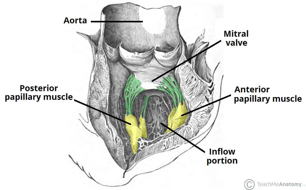
Anatomy of right ventricle. There is increasing recognition of the crucial role of the right ventricle rv in determining functional status and prognosis in multiple conditions. Pillars papillary muscles. Anterior is the largest of the three muscles.
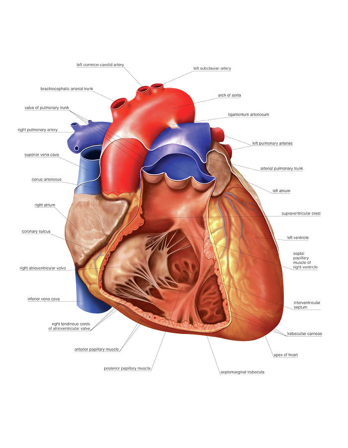
The right ventricle extends from the right atrium to the apex of the heart. Ridges attached along their entire length on one side to form ridges along the interior surface. On contrast enhanced chest ct and cardiac mri.
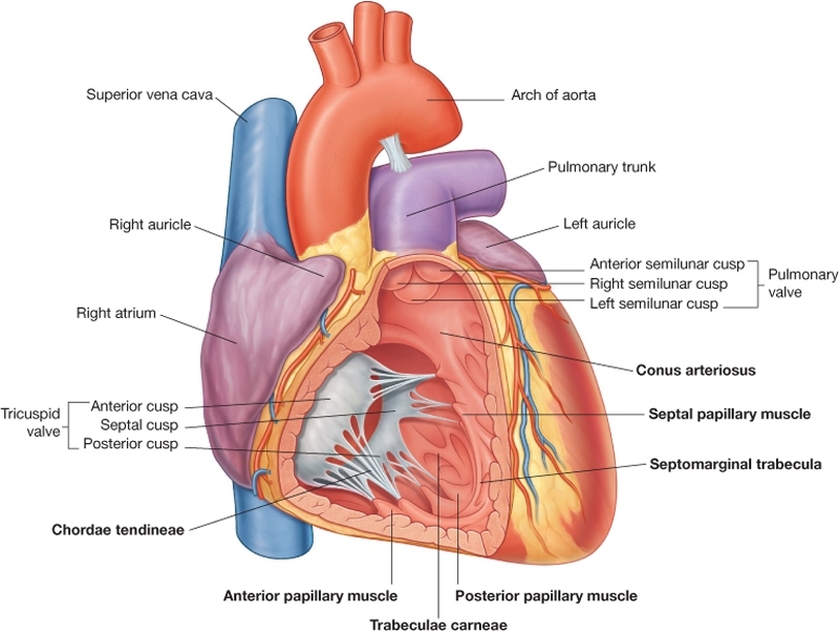
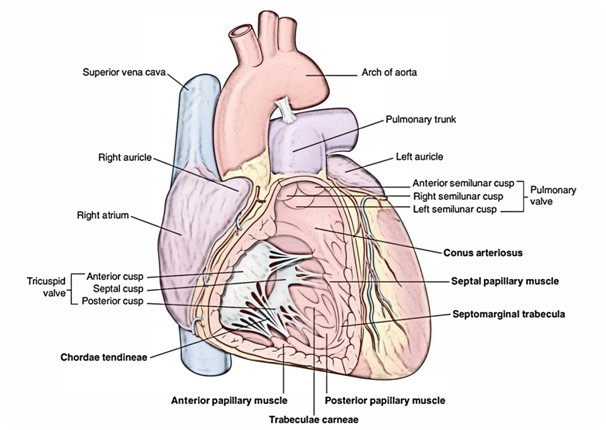
Posterior originates from the inferior wall of the right ventricle. When the ventricle contracts the right ventricle contracts the back flow of blood is prevented by the tricuspid valve which sits in this atrioventricular orifice and prevents the back flow of blood. It is located in the lower right portion of the heart below the right atrium and opposite the left ventricle.
As deoxygenated blood flows into the right atrium it passes through the tricuspid valve and into the right ventricle which pumps the blood up through. The right ventricle projects to the left of the right atrium and when viewed in. The other important internal features of the right ventricle are the papillary muscles of which there are three.
Bridges attached to the ventricle at both ends but free in the middle. Septal has attachments to the interventricular septum and. When the right atrium contracts blood is send into the right ventricle through the atrioventricular orifice.
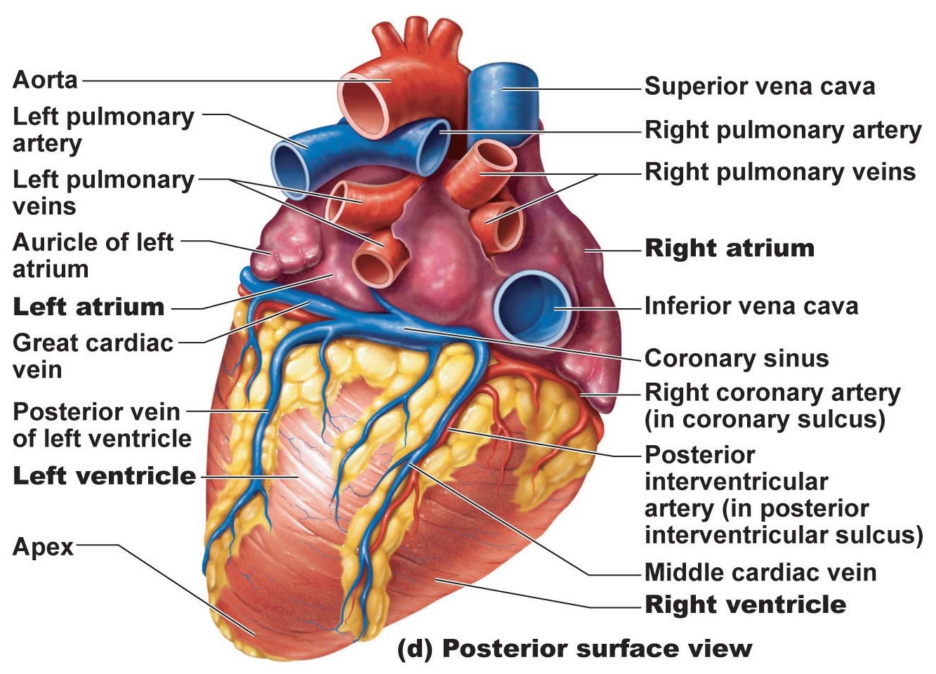
Right ventricle gross anatomy. The right ventricle pumps blood into the pulmonary circulation to the lungs and the left ventricle pumps blood into the systemic circulation through the aorta. Forms the major portion of the anterior surface of the heart.
The right ventricle in the normal heart is the most anteriorly situated cardiac chamber since it is located immediately behind the sternum. Ventricle heart in a four chambered heart such as that in humans there are two ventricles that operate in a double circulatory system. They give the ventricle a sponge like appearance and can be grouped into three main types.
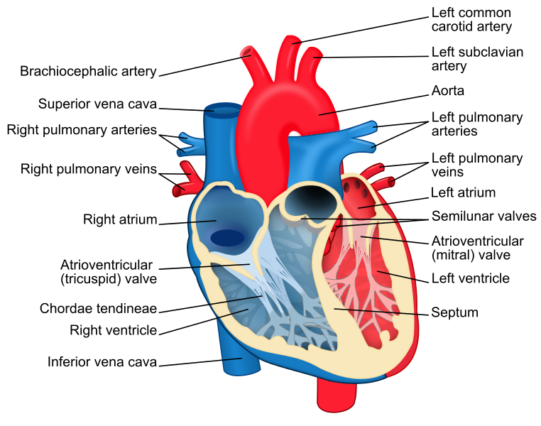
 Heart Anatomy Chambers Valves And Vessels Anatomy
 Heart Anatomy Right Ventricle 3d Anatomy Tutorial
 Chambers Of The Heart Atria Ventricles Teachmeanatomy
 World S Best Right Ventricle Stock Illustrations Getty Images
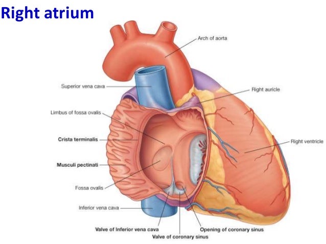
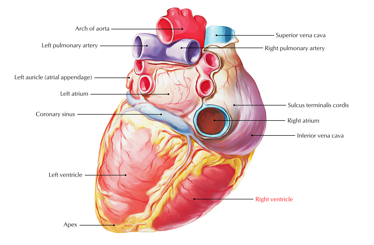
 Right Atrium And Right Ventricle
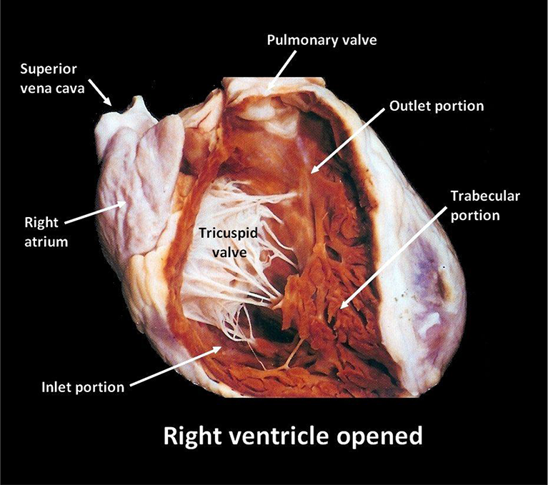
 3 Internal Features Of The Heart
Anatomy And Function Of The Coronary Arteries Johns
 Cv Physiology Cardiac Anatomy
Heart Anatomy Flashcards Memorang
 The Heart Cardiovascular System
 Anatomy Of Heart
 6 The Heart
Your Coronary Arteries Cleveland Clinic Health Information
 Easy Notes On Chambers Of The Heart Learn In Just 3
 How Your Heart Works Wake Forest Baptist Health
 Blood Flow Through The Heart Science Learning Hub
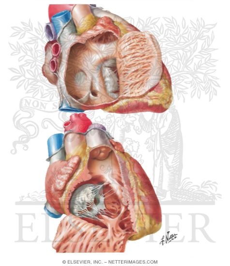
Right Ventricle Sample Netter S Anatomy Thorax Viscera
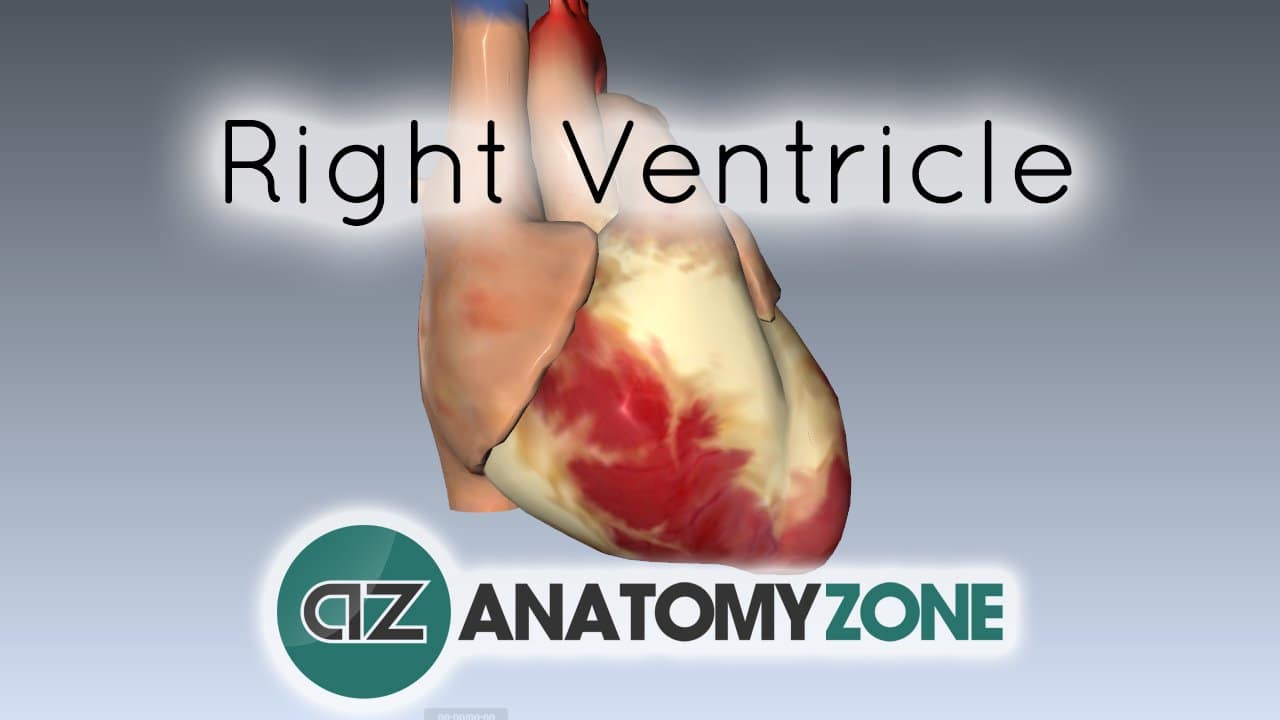 Right Ventricle Cardiovascular Anatomyzone
 Right Ventricle Earth S Lab
 Coronary Circulation Wikipedia
![]() Heart Ventricles Anatomy Function And Clinical Aspects
Heart Ventricles Anatomy Function And Clinical Aspects
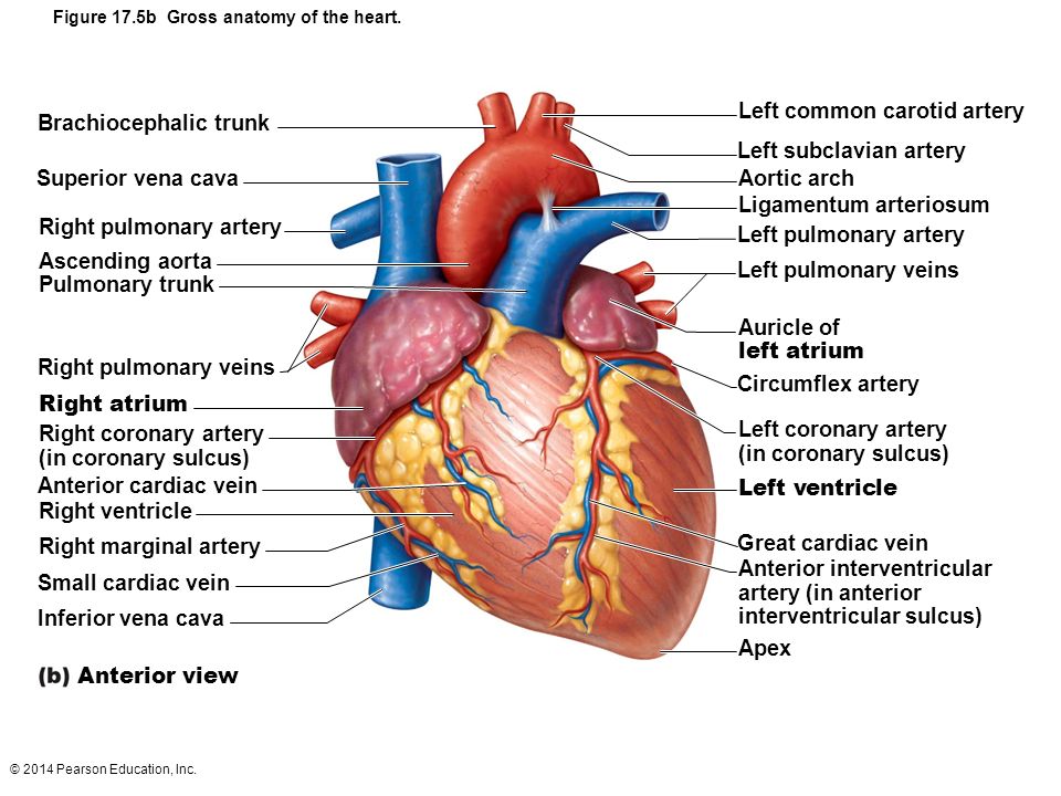 Figure 17 5b Gross Anatomy Of The Heart Ppt Video Online
 Difference Between Left And Right Ventricle Definition
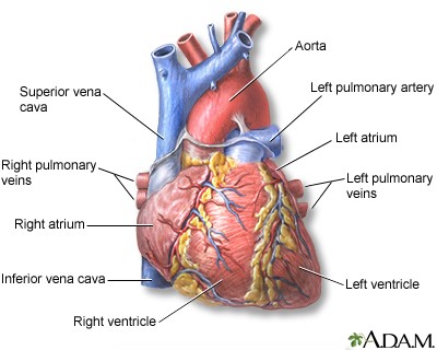
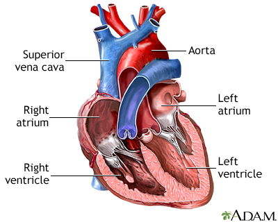 Heart Chambers Medlineplus Medical Encyclopedia Image
 Assessment Of Right Ventricle By Echocardiogram Intechopen
 Left Vs Right Ventricle Difference Between
World S Best Right Ventricle Stock Illustrations Getty Images
 Right Ventricular Infarction Part 1 Ems 12 Lead
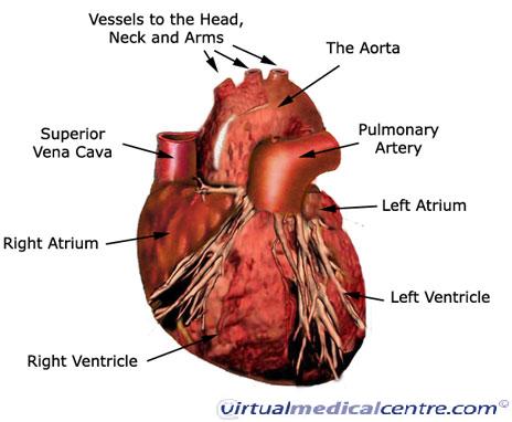 Cardiovascular System Heart Anatomy Healthengine Blog


Belum ada Komentar untuk "Anatomy Of Right Ventricle"
Posting Komentar