Anatomy Lungs And Heart
Apex the blunt superior end of the lung. Within the mediastinum the heart is separated from the other mediastinal structures by a tough membrane known as the pericardium or pericardial sac and sits in its own space called the pericardial cavity.
 Healthy Heart And Lung Anatomy
The mediastinal surface of the right lung is in contact with the heart superior vena cava inferior vena cava azygos vein and the esophagus.
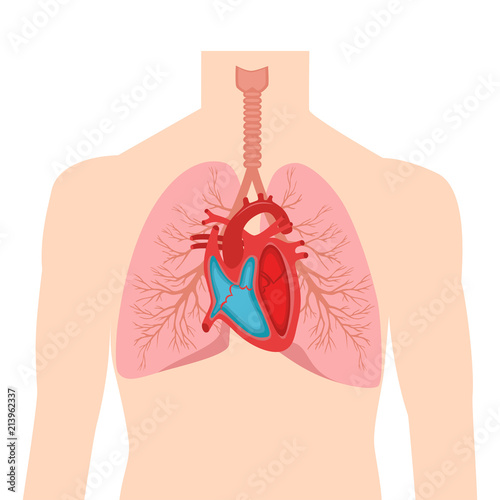
Anatomy lungs and heart. The impressions of these structures can be seen on the medial lung surface. Heart and lung anatomy this image shows the anatomy of the heart and the lungs in relation to each other displaying their different parts and features and the vessels of the heart and their relation to the lungs. The heart lies between the two lungs and is enclosed within a fibrous bag the pericardium while each lung is invested by a serous membrane the pleura.
The heart is a muscular organ about the size of a closed fist that functions as the bodys circulatory pump. Figure 1 shows the position of the heart within the thoracic cavity. The same kind of thin tissue lines the inside of the chest cavity also called pleura.
The lungs are roughly cone shaped with an apex base three surfaces and three borders. The human heart is located within the thoracic cavity medially between the lungs in the space known as the mediastinum. Here is how lungs work as the center of your breathing the path a full breath takes in your body and a 3 d model of lung anatomy.
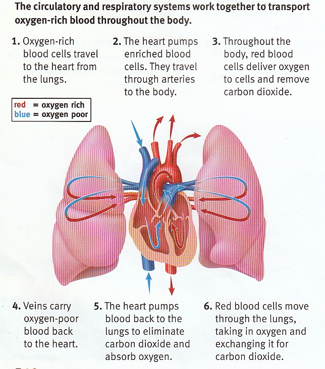
The lungs are covered by a thin tissue layer called the pleura. Each lung consists of. It takes in deoxygenated blood through the veins and delivers it to the lungs for oxygenation before pumping it into the various arteries which provide oxygen and nutrients to body tissues by transporting the blood throughout the body.
It projects upwards above the level of the 1st rib and into the floor of the neck. The lungs are the main part of your respiratory system. The heart and lungs are situated in the thorax the walls of which afford them protection.
The left lung is slightly smaller than the right this is due to the presence of the heart.
 Lungs
Lungs
 The Heart Anatomy Physiology And Function
 Anatomical Model Of Canine Heart And Lung Teaching Supplies Stem And Career Education
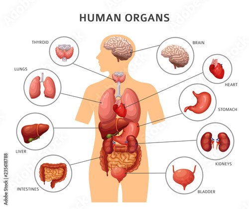
 Human Anatomy Heart Lungs Intestines Other Stock Vector
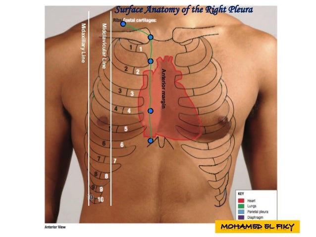 Surface Anatomy Of Heart And Lungs
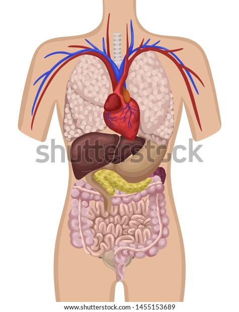
 Human Body Internal Organs Stomach And Lungs Kidneys And
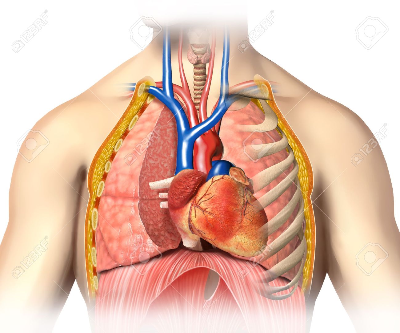 Man Anatomy Thorax Cutaway With Heart With Main Blood Veins
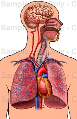
/wall-murals-human-body-anatomy-brain-lungs-heart-liver-intestines.jpg.jpg) Human Body Anatomy Brain Lungs Heart Liver Intestines Wall Mural Vinyl
Human Body Anatomy Brain Lungs Heart Liver Intestines Wall Mural Vinyl
![]() Pulmonary Arteries And Veins Anatomy And Function Kenhub
Pulmonary Arteries And Veins Anatomy And Function Kenhub
 Heart And Lung Anatomy Anterior View
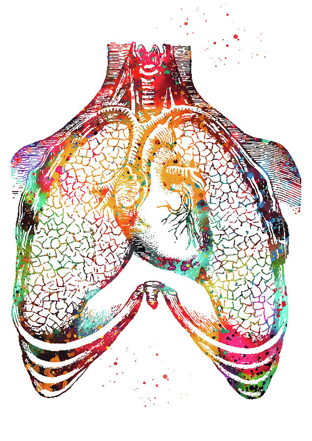
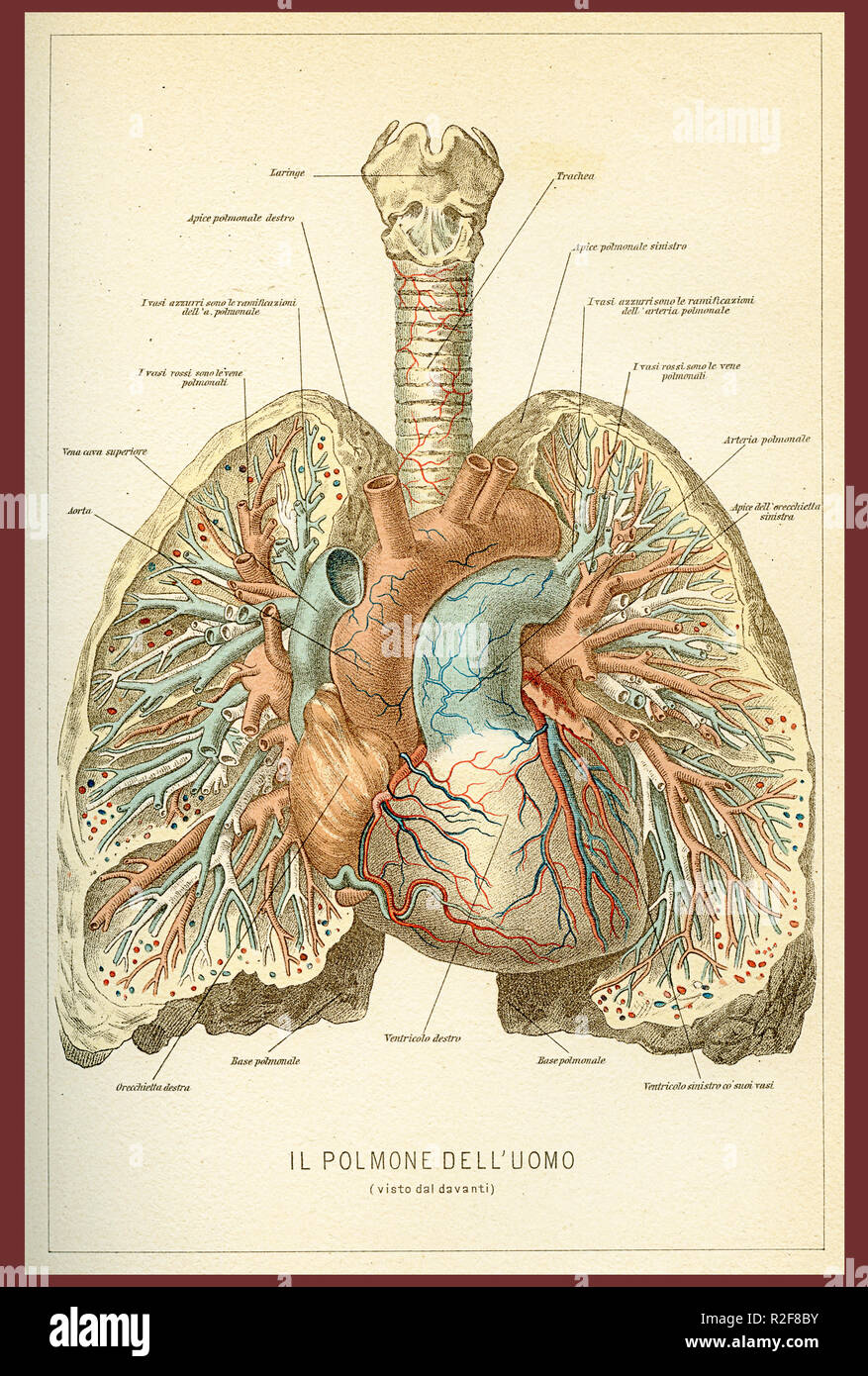 Vintage Color Table Of Anatomy Human Lungs And Heart Blood
 Lungs And Human Heart Illustration Infographic Anatomy
:max_bytes(150000):strip_icc()/heart-anatomy-581b6f483df78cc2e85bd625.jpg) How Blood Flows Through The Heart And Lungs
How Blood Flows Through The Heart And Lungs
 Lung Wikipedia
 Queensland Cardiovascular Group Anatomy Of The Heart

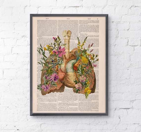 Anatomical Flower Heart Flower Lung Anatomy Art Print Christmas Gift Human Anatomy Poster Medical Art Anatomy Poster Ska099
 Heart And Lungs Internal Organs In A Male Human Body
 Anatomy Of The Heart And Great Vessels Medical

 Newsela Why Do You Have Two Lungs But Only One Heart
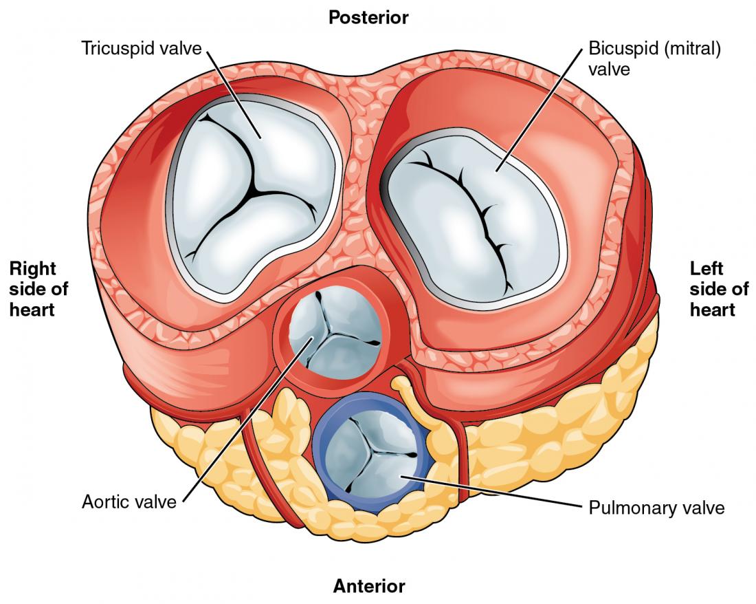
 Heart Anatomy Yourheartvalve
 Human Heart And Lungs
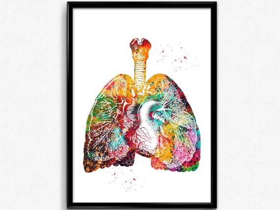
 Heart And Lungs Anatomy
 Heart Anatomy Anatomy And Physiology
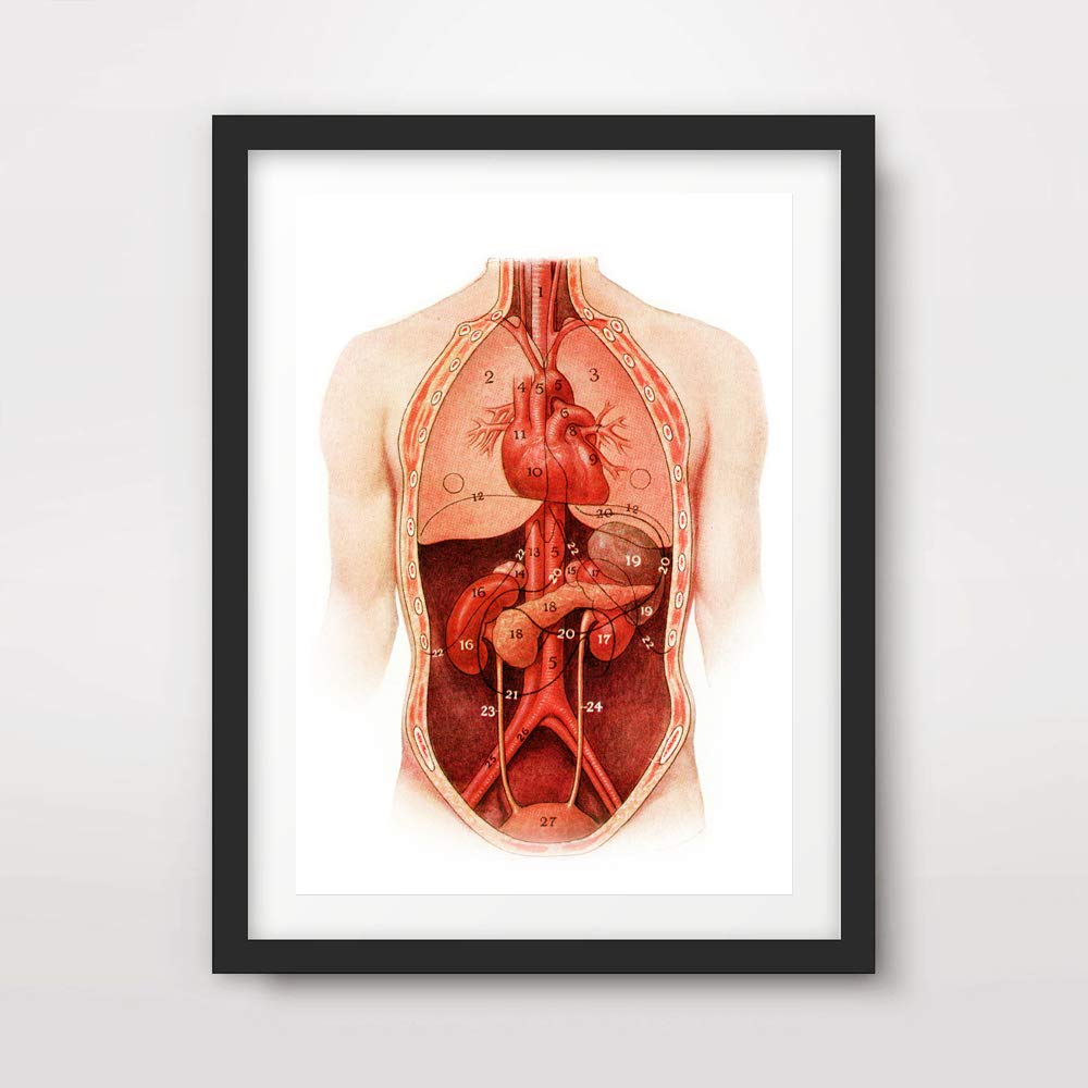
Amazon Com Heart Lungs Kidneys Medical Art Print Anatomical
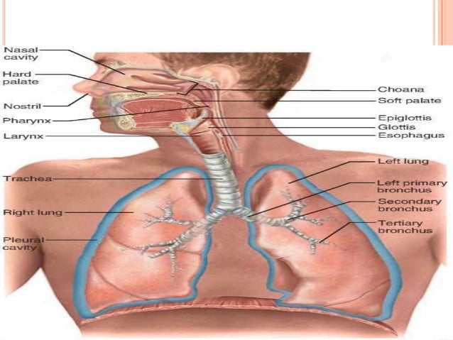
 Chest Anatomy Heart And Lungs
 Canine Heart And Lung Model
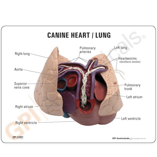 Anatomical Model Canine Heart And Lung
 Anatomy And Physiology Of Heart Lung
 Lungs And Heart Anatomy Art Print Cardiovascular System Poster Anatomy Art Physiology Medical Art Poster 1236
 Model Human Body With Liver Kidney Lungs And Heart Stock
Belum ada Komentar untuk "Anatomy Lungs And Heart"
Posting Komentar