Anatomy Of Spleen
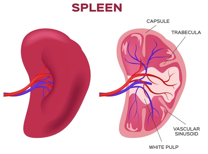
It is wrapped by a fibroelastic capsule which allows the spleen to significantly increase its size when necessary. By pressing on the belly under the left ribcage.
 Anatomy Of Spleen And It S Applied Aspect
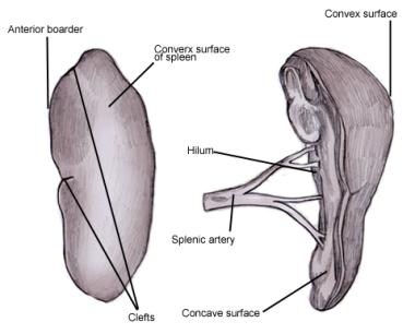
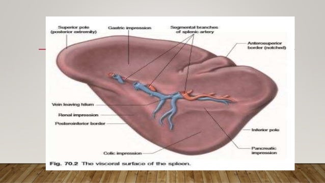
The anterior end of the spleen is expanded and is more like a border.

Anatomy of spleen. A probe is placed on the belly and harmless sound waves create images by reflecting. It rests on the upper pole of the left kidney. Spleen tests physical examination.
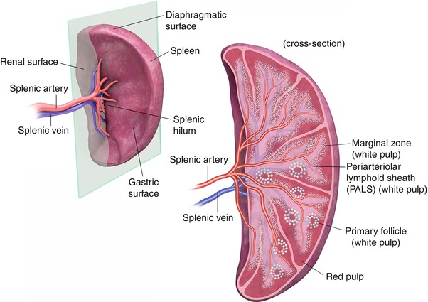
It is the largest organ in the bodys lymphatic system which is responsible for promoting immune function filtering the blood and managing blood volume. The spleens 2 ends are the anterior and posterior end. The spleen is a soft organ with a thin outer covering of tough connective tissue called a capsule.
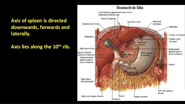
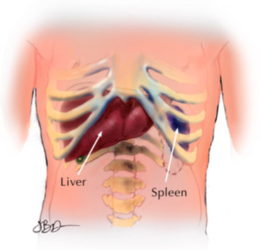
Spleen organ of the lymphatic system located in the left side of the abdominal cavity under the diaphragm the muscular partition between the abdomen and the chest. The spleen is a small organ typically located on the left side of the body behind the ribcage and stomach. The spleen is an intraperitoneal organ so all of its surfaces are covered with visceral peritoneum.
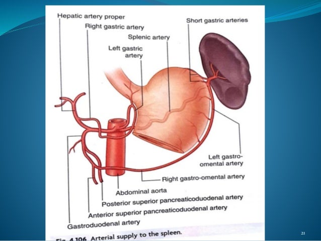
While the bone marrow is the primary site of hematopoiesis in. The spleen is rich in blood supplied via the splenic artery. Production of opsonins properdin and tuftsin.
The spleen is connected to the stomach and kidney by parts of the greater omentum a double fold of peritoneum that originates from the stomach. Computed tomography ct scan. Other functions of the spleen are less prominent especially in the healthy adult.
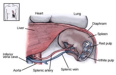
Splenorenal ligament posterior to. It is directed forward and downward to reach the midaxillary line. The spleen is a purple fist sized organ.
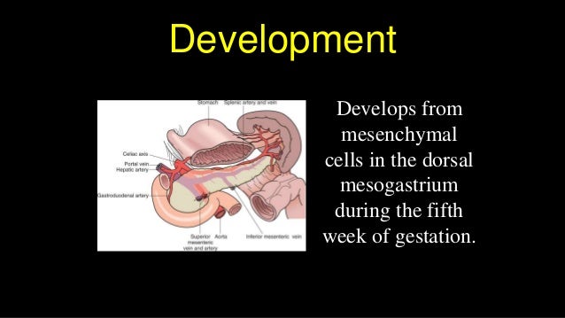
The posterior end is rounded and is directed upward and backward. Spleen produces all types of blood cells during fetal life. There is a handy rule to remember the rough dimensions of the spleen called the 1x3x5x7x9x11.
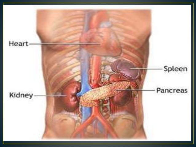
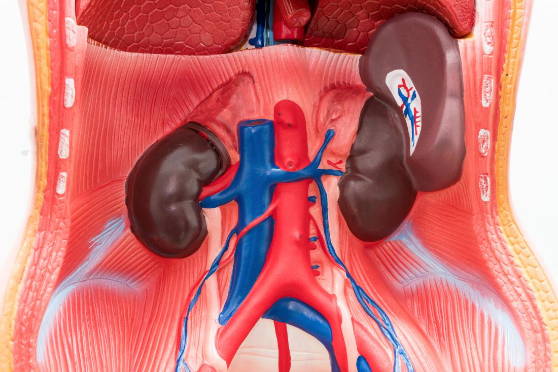
In humans it is about the size of a fist and is well supplied with blood. It is positioned under the rib cage below the diaphragm and above the left kidney. Creation of red blood cells.
Gastrosplenic ligament anterior to the splenic hilum connects the spleen to the greater curvature of the stomach. Blood exits this organ through the splenic vein. Magnetic resonance imaging mri.
A ct scanner takes multiple x rays. The spleen also contains efferent lymphatic vessels which transport lymph away from the spleen.
![]() Spleen Anatomy Location And Functions Kenhub
Spleen Anatomy Location And Functions Kenhub
 Differences In Tissue Anatomy Characteristics Between Nmr
 Anatomy And Physiology The Adventures Of Super Spleen
 Spleen Anatomy
 The Spleen Anatomy Function And Disease
![]() Spleen Anatomy Location And Functions Kenhub
Spleen Anatomy Location And Functions Kenhub
 Dictionary Normal Spleen The Human Protein Atlas
 Amazon Com Liver Stomach Pancreas Gallbladder Spleen
 Cross Sectional Imaging Of The Spleen Radiology Key
 Anatomy Of Splenectomy For Ruptured Spleen Sciencedirect
 Living Without A Spleen
 Anatomy Of Spleen
 Arteries Of The Pancreas Duodenum And Spleen Preview Human Anatomy Kenhub
 Spleen Anatomy Overview Gross Anatomy Microscopic Anatomy
 Spleen Anatomy 3d Medical Vector Illustration
![]() Spleen Anatomy Icon Cartoon Style
Spleen Anatomy Icon Cartoon Style
 Spleen Anatomy Organs Body Organs Diagram Human Anatomy
![]() Spleen Anatomy Icon Cartoon Style Stock Vector Art
Spleen Anatomy Icon Cartoon Style Stock Vector Art
 Anatomy Of Spleen

 Anatomy Of Spleen
![]() Spleen Anatomy Location And Functions Kenhub
Spleen Anatomy Location And Functions Kenhub
 Spleen Anatomy And Physiology
 Spleen Anatomy Overview Gross Anatomy Microscopic Anatomy
 Anatomy Of The Pancreas And Spleen Sciencedirect
 Anatomy Of Spleen
 What Is The Spleen Cll Society
 Anatomy Of The Pancreas And Spleen Sciencedirect
Spleen
Belum ada Komentar untuk "Anatomy Of Spleen"
Posting Komentar