Shins Anatomy
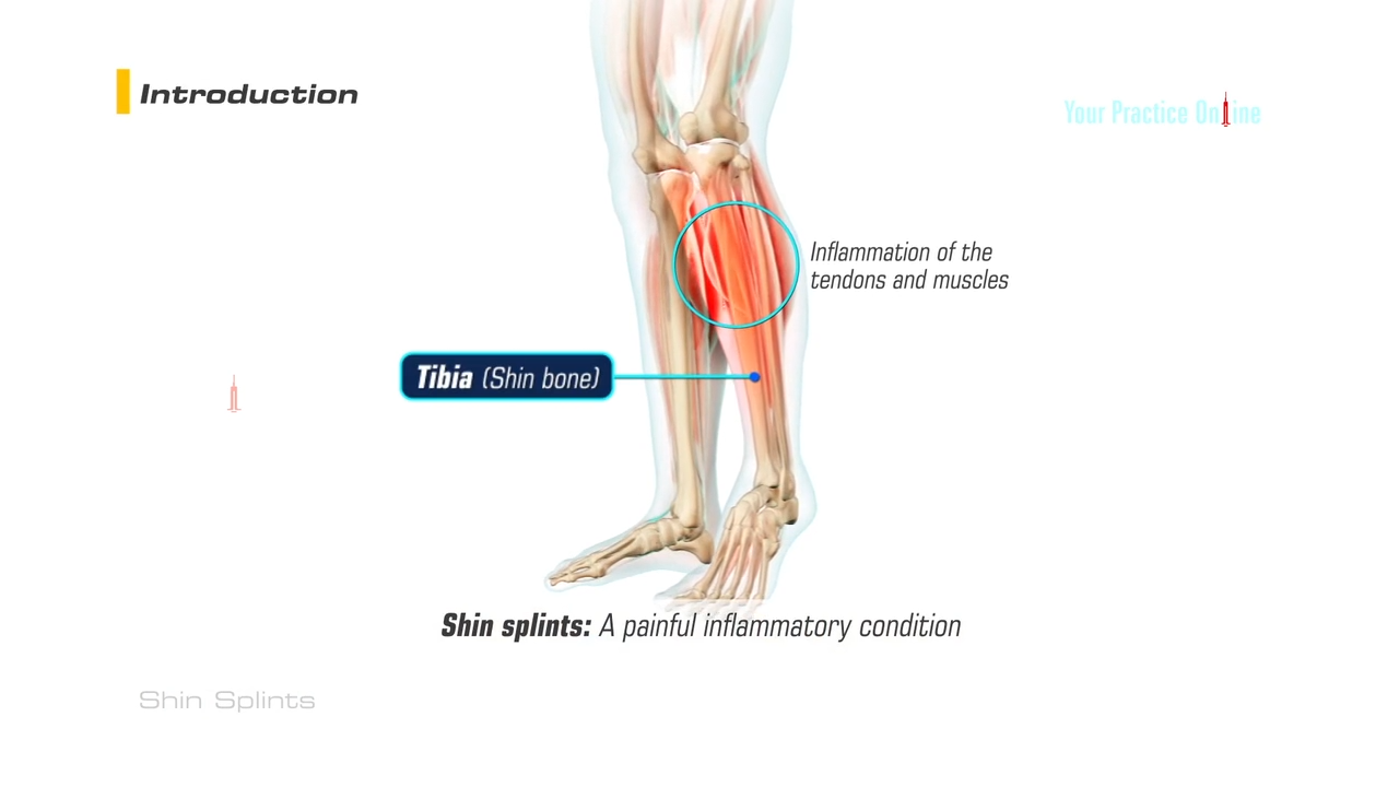
Shin splints medial tibial stress syndrome is an inflammation of the muscles tendons and bone tissue around your tibia. Anatomy of shin splints.
 Tibia Bone Anatomy Pictures Definition Body Maps
Shin splints frequently affect people who engage in moderate to heavy physical activity.

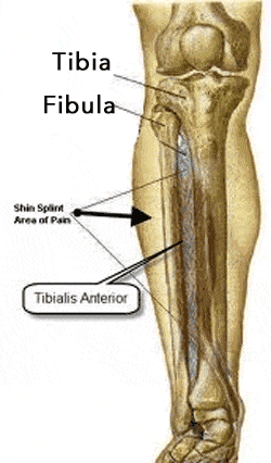
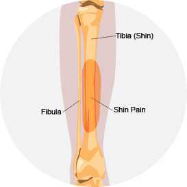
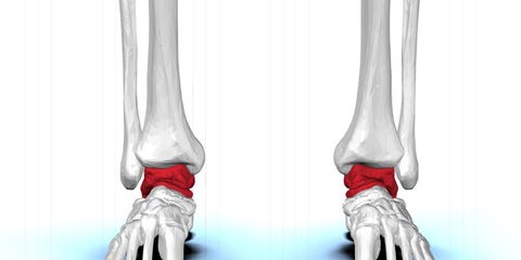
Shins anatomy. Shin splint pain most often occurs on the inside edge of your tibia shinbone. The deep posterior compartment. There are two bones in the shin area.
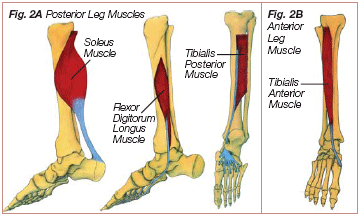
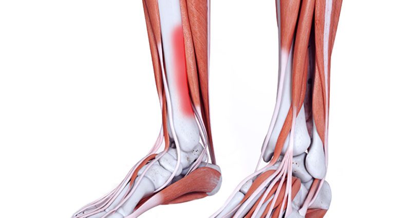
With the popularity of muay thai in mma we are taught to throw and block roundhouse kicks with our shins. Shin muscles such as the tibialis anterior and extensor digitorum longus dorsiflex the foot and extend the toes. The muscles of the lower leg the anterior compartment in the front of the shin holds the tibialis anterior.
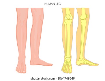
The tibia is also known as the shinbone and is the second largest bone in the body. In general shin splints develop when the muscle and bone tissue periosteum. Cookery chiefly brit a cut of beef the lower foreleg.
Definitiondescription medial tibial stress syndrome mtss or shin splint syndrome is a clinical pain condition defined as exercise induced pain along the posteromedial tibial border distal third caused by repetitive loading stress during running and jumping and provoked on palpation over a length of 5consecutive centimeters. The posterior compartment holds the large muscles that we know as the calf muscles. Shin splints medial tibial stress syndrome is an inflammation of the muscles.
Your doctor may refer to the condition as medial tibial stress syndrome mtss. The tibia and fibula or calf bone. These two bones connect the ankle to.
The most common symptom of shin splints is pain. Pain typically occurs along the inner border of the tibia where muscles attach to the bone. The term shin splints describes pain felt along the front of your lower leg at the shin bone.
Anatomy the front edge of the tibia. Anatomy the front part of the lower leg. The lateral compartment is along the outside of the lower leg.
The muscles of the calf also work subtly to stabilize the ankle joint and foot and to maintain the bodys balance. The fibula is smaller and thinner than the tibia. Treatment and prevention description.
This pain concentrates in the lower leg between the knee and ankle. But the shin bone or tibia is a huge bone that varies in size and shape. The tibia is a large bone located in the lower front portion of the leg.
 Shin Splints Orthogate
 Shin Splints
 Shin Splints Powerpoint
Shin Splints Powerpoint
 Caspianrehab Treatment For Shin Splint Do Your Shins
 Banish Shin Splints Forever With One Magical Exercise Shin
 Getting A Leg Up On Shin Pain Dubin Chiropractic
 Bluegrass Foot Centers Podiatry
Pin On Health And Exercise
 Tibialis Anterior Muscle Wikipedia
 Shin Splints Wikipedia
:max_bytes(150000):strip_icc()/GettyImages-149678870-5702c33f5f9b581408a76e0b.jpg) The Anatomy Of The Lower Leg Muscles
The Anatomy Of The Lower Leg Muscles
 Medial Tibial Stress Syndrome Part 1 What Are Shin Splints
 Medial Tibial Stress Syndrome Part 1 What Are Shin Splints
 Summit Medical Group
 Shins And Calves Back In The Game Physical Therapy
 Tibia Anatomy And Function
 My Dirt Road Anthem Shin Pain Not Good
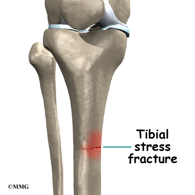
Tibia Shinbone Shaft Fractures Orthoinfo Aaos
 Why Doing Shin Exercises Won T Help You Avoid Shin Splints
 The Lower Leg The Calf And The Shin
 Shin Splints 321gomd
 Shin Splint Treatment In Nyc Nydnrehab Com
 Tibialis Anterior Muscle Functional Anatomy Guide
Tibia Shinbone Shaft Fractures Orthoinfo Aaos
 Shin Splints Leg Pain Salt Lake City Ut Foot Doctor
Shin Splints Orthoinfo Aaos
 Names Of Nerves In Hands Shins And Face Biology Stack
 Shin Splints Symptoms Causes Treatment Taping Exercises
 Why Does My Leg Hurt Shin Splints Tendinitis Stress
 Shin Splint Treatment In Nyc Nydnrehab Com
 Medial Tibial Stress Syndrome An Overview Sciencedirect
 Anatomy Of Shin Splints Treatment And Prevention
 Royalty Free Shin Bone Stock Images Photos Vectors


Belum ada Komentar untuk "Shins Anatomy"
Posting Komentar