Airway Intubation Anatomy
A keen understanding of airway anatomy can make the process of intubating a patient much easier. It is frequently performed in critically injured ill or anesthetized patients to facilitate ventilation of the lungs including mechanical ventilation and to prevent the possibility of asphyxiation or airway obstruction.
 Rt 127
Robert bastian provides an introduction to larynx pharynx and airway anatomy.
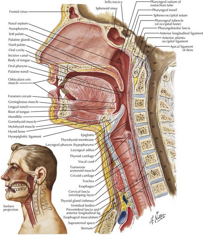
Airway intubation anatomy. Paediatric airway anatomy appropriately positioning children undergoing intubation. Formed by union of facial bones nasal floor towards ear not eye lined with mucous membranes cilia tissues are delicate vascular adenoids. The nasal fossae are divided by the midline cartilaginous septum and medial portions of the lateral cartilages fig.
Warm filter and humidify air. Knowledge of the functional anatomy of the airway in these forms the basis of understanding the pathological conditions that may occur. Selecting the correct equipment for intubation.
Try using search on phones and tablets. Tracheal intubation usually simply referred to as intubation is the placement of a flexible plastic tube into the trachea to maintain an open airway or to serve as a conduit through which to administer certain drugs. Navigation best viewed on larger screens.
This information is meant to serve as an educational resource. Endotracheal intubation is a basic skill that every first responder must master. Lymph tissue filters bacteria commonly infected.
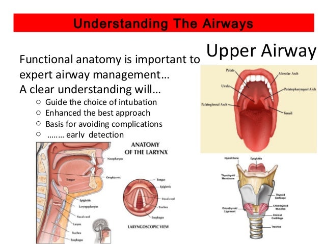
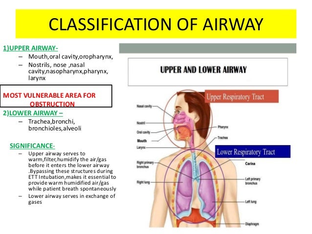
Interpreting structures seen on direct laryngoscopy. The upper airway extends from the mouth to the trachea. And while it does pose some risks it is also safe with the right technique and diligent attention to the patient.
The two nasal fossae extend from the nostrils to the nasopharynx. Nasotracheal intubation is an alternative approach to orotracheal intubation. This demonstration by anthony lewis from isimulate and todd slesinger provides a brief overview of the basics of the upper airway and laryngoscopy.
A good understanding of airway and intubation is fundamental to managing a sick patient. Home airway and intubation. It includes the mouth the nose the palate the uvula the pharynx and the larynx.
The most widely used route is orotracheal in which an en. In this presentation dr. Nasal cavity and nasopharynx.
 Regional And Topical Anesthesia For Awake Endotracheal
 Medications Used In Tracheal Intubation Medications For
 Pin On Intubation
 Paediatric Airway Anatomy Paediatric Emergencies
 Tracheal Intubation Wikipedia
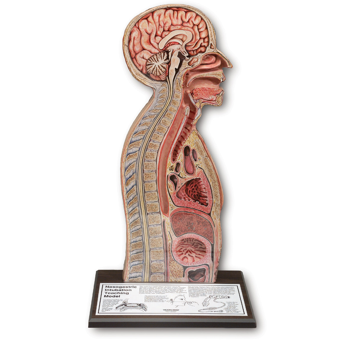 Nasogastric Intubation Model
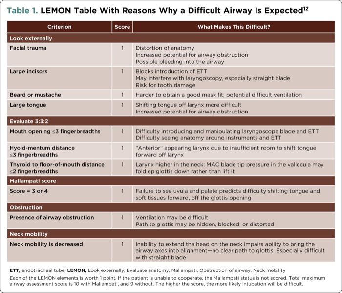
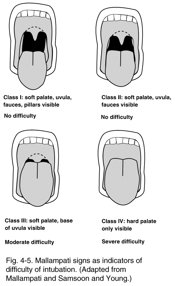
 Airway Management Respiratory Tract Mallampati Score

 Anatomical Differences Between Pediatric And Adult Airways
 Ultrasound Gel
 Airway Intubation Stock Photos Airway Intubation Stock


 Airway Management Outline Review Of Airway Anatomy Airway
 Tracheal Intubation Critical Care Medicine Merck Manuals
:max_bytes(150000):strip_icc()/GettyImages-188057983-625125a51be848dfae5fbfa219f5a6f7.jpg) What Is Intubation And Why Is It Done
What Is Intubation And Why Is It Done
 Chapter 38 Airway Management Principles And Practice Of
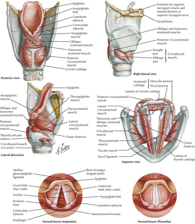 Tracheal Intubation And Endoscopic Anatomy Basicmedical Key
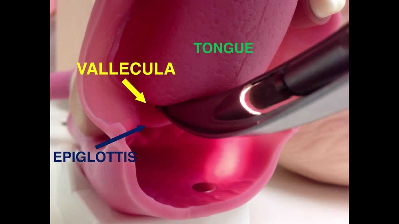 Airway Devices 01 Direct Laryngoscopy
 Upper Airway Assessment East Iv
 Airway Anatomy Its Assessment And Anaesthetic Implication
 Figure 1 From Airway Regional Anesthesia For Awake
 10 Rules For Approaching Difficult Intubation
 Chapter 122 Intubation And Airway Support Principles And
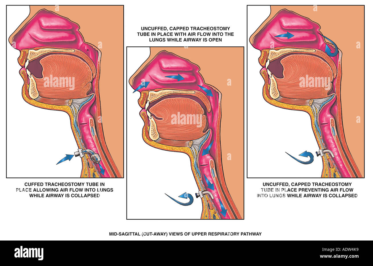
 Intubation And Tracheostomy Medical Illustration Human
 Functional Anatomy And Physiology Of Airway Intechopen
 Tracheal Intubation And Endoscopic Anatomy Basicmedical Key

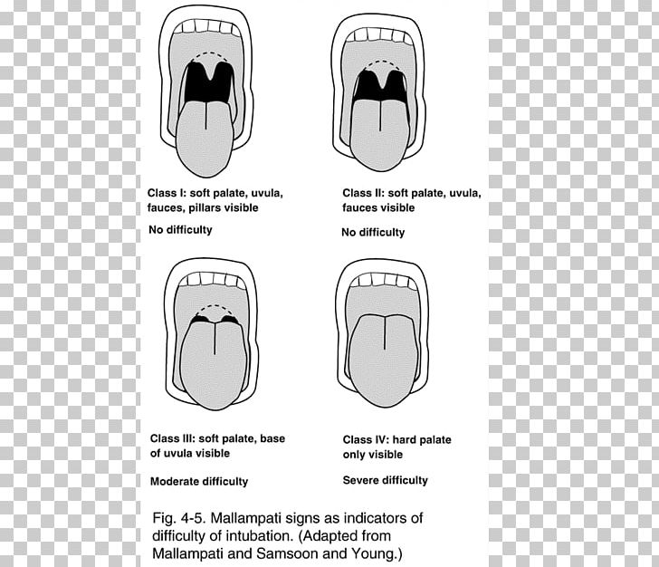 Airway Management Respiratory Tract Mallampati Score
 Emergency Airway Management
 Intubation Step By Step Intubation Technique Explained
 Functional Anatomy And Physiology Of Airway Intechopen
 Airway Skills 3 Orotracheal Intubation
10 Rules For Approaching Difficult Intubation
Belum ada Komentar untuk "Airway Intubation Anatomy"
Posting Komentar