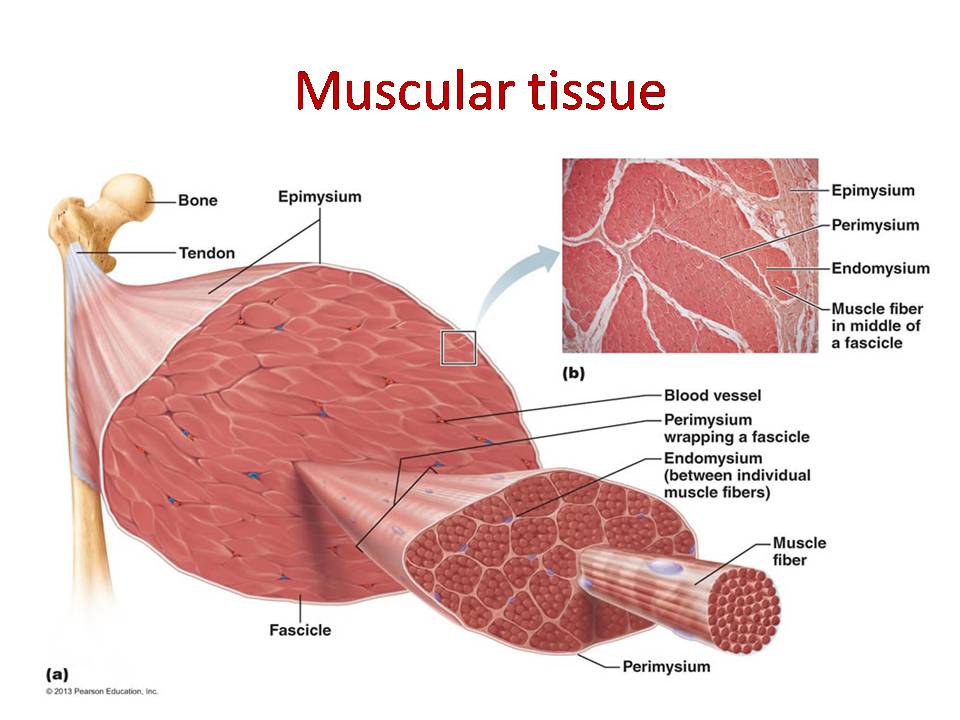
Muscle Cell Anatomy
These are the nucleuses. A muscle cell known technically as a myocyte.
 Intercalated Disc Cardiac Muscle Gap Junction Muscle Tissue
And then if you take the cross section of that there are tubes within that called myofibrils.

Muscle cell anatomy. Which qualities of a skeletal muscle fiber can change as a person ages. And again this endomysium just like the perimysium contains nerves and blood vessels that can help conduct neuronal signals and blood towards the individual myofiber and the connective tissue that sits around here. Muscle cell muscle cell definition.
Striations indicate that a muscle cell is very strong unlike visceral muscles. Structure of a muscle cell. Why is an individual skeletal muscle cell referred to as a muscle fiber.
The arrangement of protein fibers inside of the cells causes these light and dark bands. Because of its thread like shape. Muscle tissue one of the four major tissue types plays the vital role of providing movement and heat generation to the organs of the body.
So we can call it a muscle cell but we can also call it a myo myo meaning muscle fiber. The cells of cardiac muscle tissue are striatedthat is they appear to have light and dark stripes when viewed under a light microscope. Each nucleus regulates the metabolic.
Within muscle tissue are three distinct groups of tissues. Smooth muscle or involuntary muscle is found within the walls of organs and structures such as. There are three types of muscle tissue recognized in vertebrates.
The mechanism of muscle contraction. Function of a muscle cell. The membrane of the muscle cell is the sarcolemma.
So this is shaped like a fiber because it is longer than it is wide. Anatomy test 5152 62 terms. Skeletal muscle or voluntary muscle is anchored by tendons or by aponeuroses at a few places.
Muscle cells rarely act alone muscle organs operate on principle of graded strength motor units the functional unit of muscle system motor unit individual motor neuron and all muscle cells that it innervates the axon of a motor neuron usually branches on entering a muscle bundle and a single axon may innervate a few to 100s of muscle. Cardiac muscle myocardium. Skeletal muscle cells muscle cells commonly known as myocytes are the cells that make up muscle tissue.
Skeletal muscle cells are long cylindrical multi nucleated and striated. Each of these tissue groups is made of specialized cells that give the tissue its unique properties. There are 3 types of muscle cells in the human body.
As seen in the image below a muscle cell is a compact bundle. Certain heart defects. Skeletal muscle cardiac muscle and smooth muscle.
Sarcomeres action potential and the neuromuscular junction duration. Professor dave explains 89683 views. To activate a muscle the brain sends an impulse down a nerve.
Anatomy and physiology teas science prenursingsmarterpro teas guide.
 Anatomy Of A Skeletal Muscle Cell Video Khan Academy
 Smooth Muscle Cell Vector Anatomy Relaxed And Contracted
 10 2 Skeletal Muscle Anatomy And Physiology
 Levels Of Organization For The Muscular System Lessons
 Muscle Cell Anatomy Physiology A Review Quiz Worksheet
 2014 Pearson Education Inc Human Anatomy Skeletal Muscle
 Muscular Tissue Skeletal Smooth And Cardiac Muscle
 Ppt Chapter 6 The Muscular System Part A Powerpoint
 Muscular System Muscles Of The Human Body
 Microscopic Anatomy And Organization Of Skeletal Muscle
 Skeletal Muscle Cell Anatomy Amp Physiology Pdf Skeletal
 Anatomical Review Of Muscle Cell Anatomy Fig Whole Muscle
 Muscle Cell Anatomy Physiology A Review Quiz Worksheet
 Muscle Biology Physiology Basic Science Orthobullets
 Exercise 14 Microscopic Anatomy And Organization Of Skeletal
 The Anatomy Of A Muscle Cell W Key
 The Muscular System How We Move Around Interactive
 Muscle Fiber Diagram Muscle Fiber Cell Myofibril
 Image Result For Skeletal Muscle Cells Diagram With
 Leg Muscle Anatomy High Impact Visual Litigation Strategies
 10 2 Skeletal Muscle Anatomy Physiology


Belum ada Komentar untuk "Muscle Cell Anatomy"
Posting Komentar