Anatomy Of The Talus
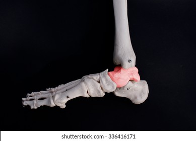
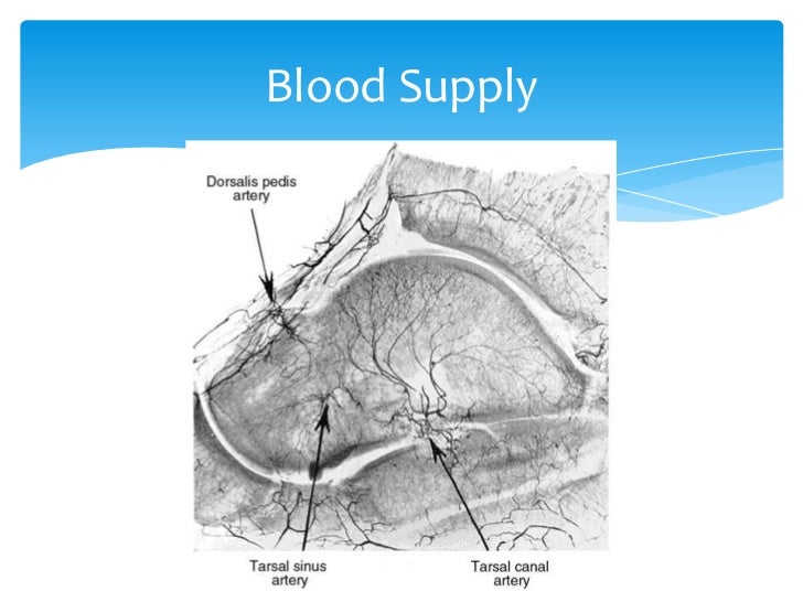
The regions supplied by the three arteries that vascularize the talus are highlighted and labeled. The talus bone is the bone that connects the lower leg bones to the foot.
 Osteochondral Lesions Of The Talus Background Anatomy
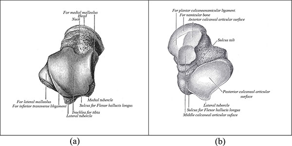
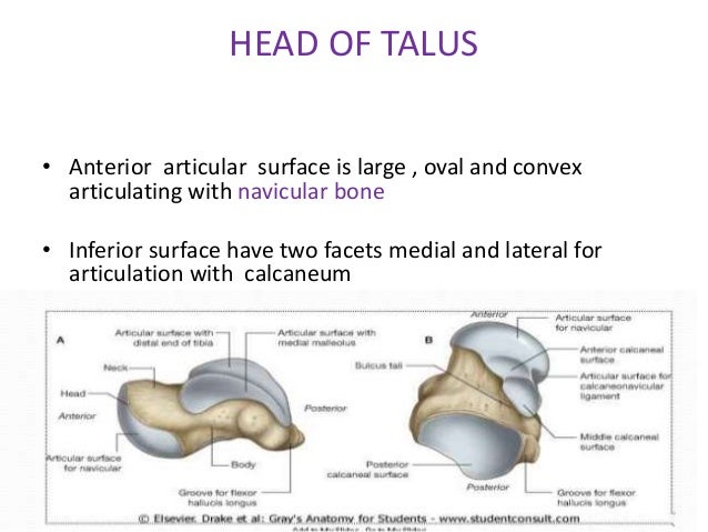
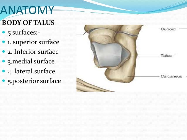
It presents with five surfaces.

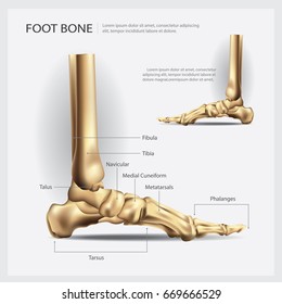
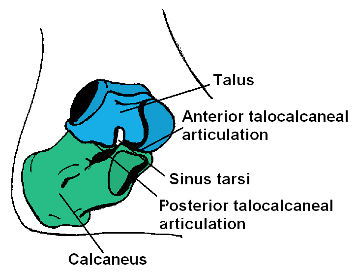
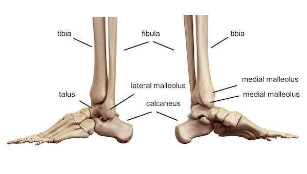
Anatomy of the talus. The talus is a tarsal bone in the hindfoot that articulates with the tibia fibula calcaneus and navicular bones. The talus is an important bone of the ankle joint that is located between the calcaneus heel bone and the fibula and tibia in the lower leg. The os trigonum is a normal variant of talar anatomy representing an unfused lateral tubercle of the posterior process.
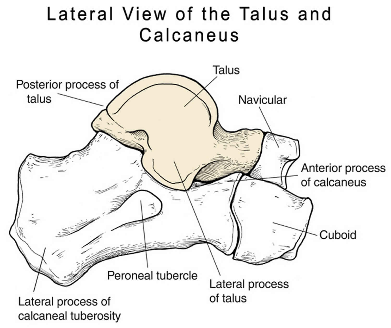
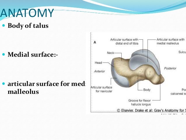
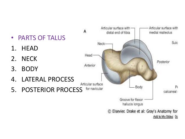
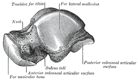
The main anatomic landmarks of the talus are indicated. A superior inferior medial lateral and a posterior. Muscle and ligamentous attachments.
Ankle fractures are often fractures of the talus. The transverse diameter of the body is greater anteriorly than posteriorly. The groove on the posterior surface lodges the tendon of the flexor hallucis longus.
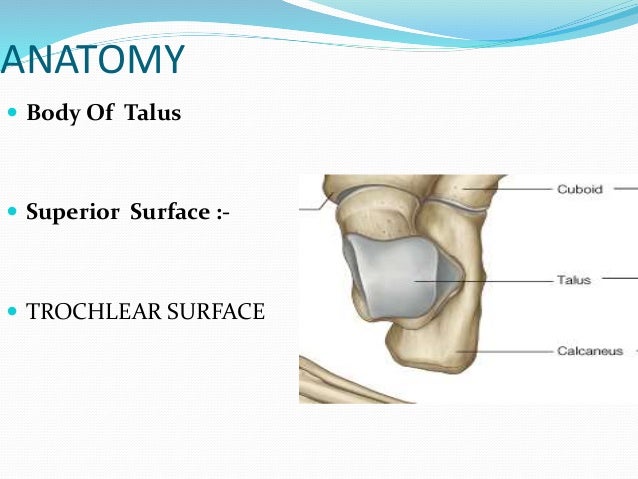
7 the superior surface of the body presents behind a smooth trochlear surface the trochlea for articulation with the tibia. 1 the dome or body of the talus articulates with the tibia and fibula on its superior medial and lateral surfaces to form the ankle joint. The talus is pivotal to the function of the ankle literally.
The top of the talus contains round cradle like depressions that the lower leg bones fit into. Anatomy of the talus. No muscles are attached.
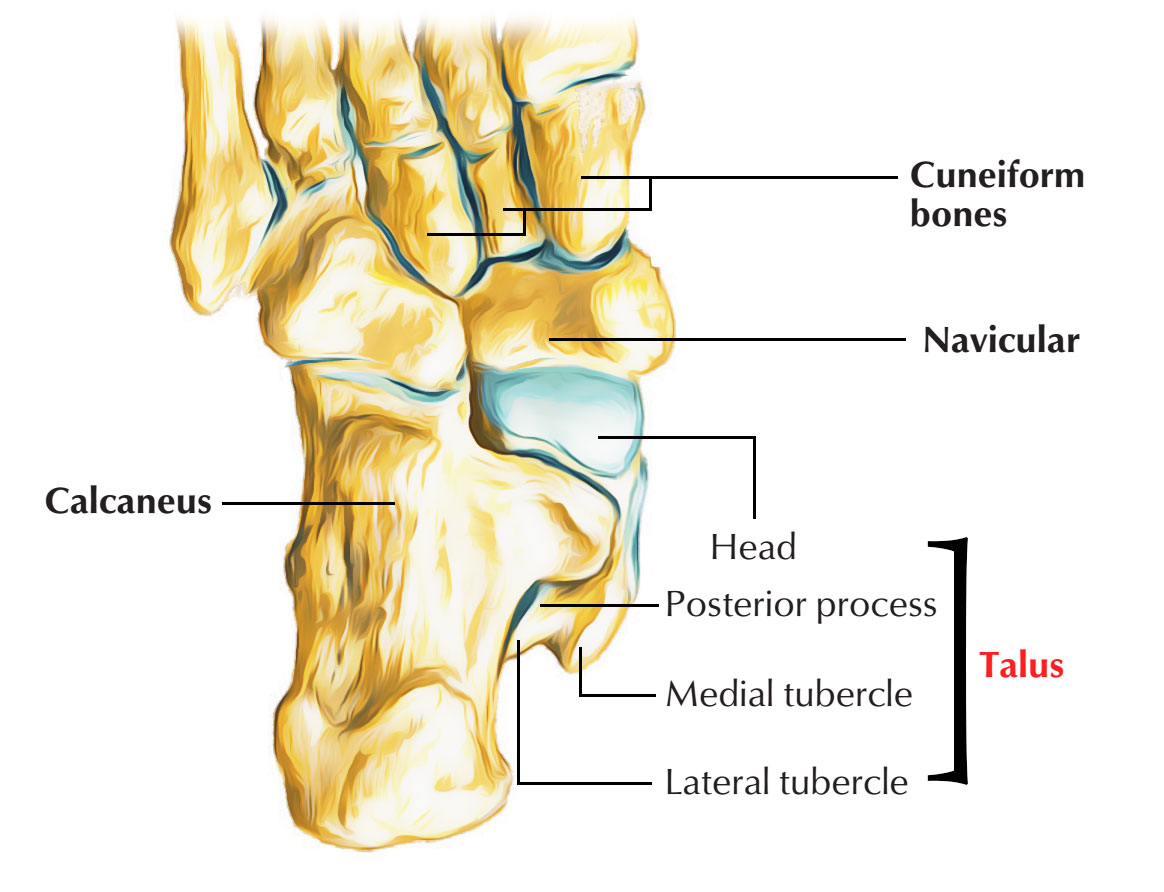
The shape of the bone is irregular somewhat. The head of the talus has a convex surface and carries the articular surface of the navicular bone. Anatomy and blood supply.
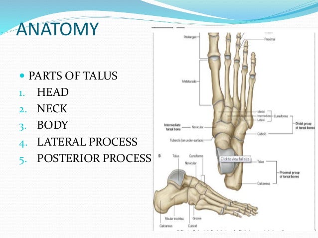
The talus is a very compact and hard bone making up a part of the ankle joint where. The talus is a uniquely shaped bone divided into three anatomic regions. The medial tubercle provides attachment to the superficial fibers of the.
These bones rotate within. It has no muscular attachments and around 60 of its surface is covered by articular cartilage. The body of the talus comprises most of the volume of the talus bone ankle bone.
The talus is part of a group of bones in the foot which are collectively referred to as the tarsus. The topmost bone of the foot anatomy. Body of talus the lower non articular part of the medial surface of the body gives attachment to the deep fibers of the deltoid ligament.
 Tarsus Anatomy Of The Dog On Ct
 Talus Bone Images Stock Photos Vectors Shutterstock
 Anatomy Ct Images Volume Rendering Technology Vrt Of
 Talus Fracture
 Talus Fracture Causes Types Symptoms Complications
 Talus Bone Images Stock Photos Vectors Shutterstock
 Talus Bone Wikipedia
![]() Talus Anatomy And Clinical Aspects Kenhub
Talus Anatomy And Clinical Aspects Kenhub
 Talus Fracture
 The Diagnosis Management And Complications Associated With
 Talus
Talus
 Talus
Talus

 Ankle Foot Anatomy
 Talus Fracture

 Ankle Joint An Overview Sciencedirect Topics
 Easy Notes On Talus Learn In Just 4 Minutes Earth S Lab

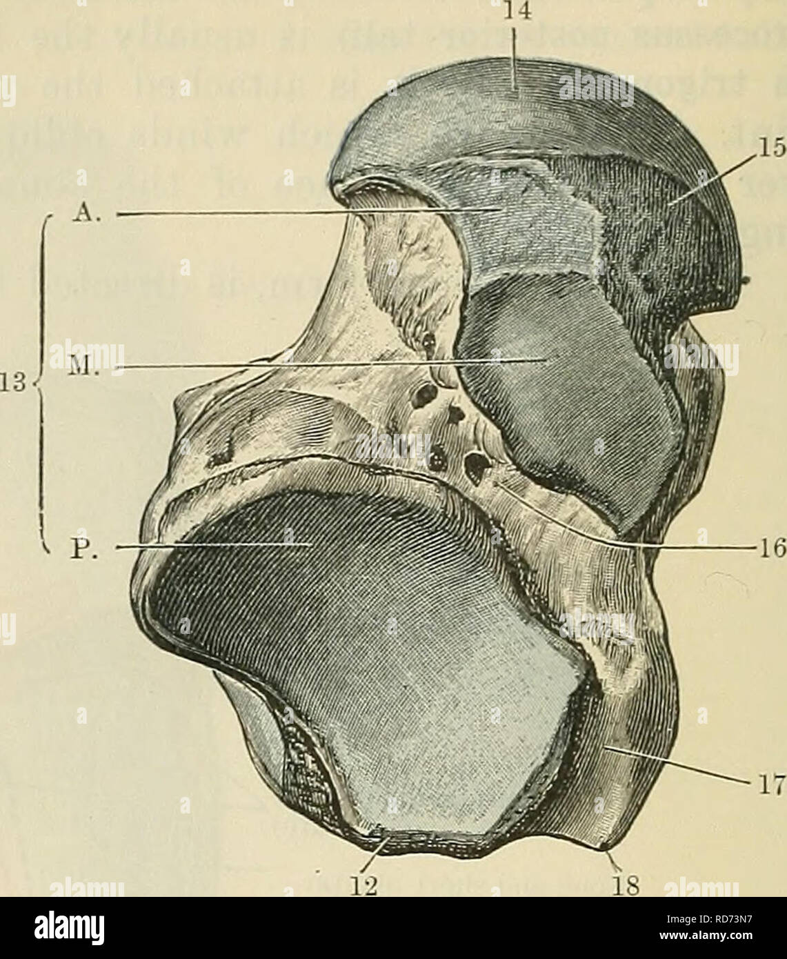 Cunningham S Text Book Of Anatomy Anatomy Fig 257 The
 Talus Fracture
 Ecr 2006 C 509 Fractures Of The Talus A Pictorial
 Talus Fracture Other Than Neck Trauma Orthobullets
 Talus Bone Wikipedia
 Anatomy Of The Talus Radiology Case Radiopaedia Org
Get To Know The Ankle Joint Yoga Journal
 Talus Anatomy Blood Supply Fractures
 Diagram Talus Calcaneus Articulation Anatomy Diagram
 Talus Bone Anatomy Bone And Spine
 Talus Radiology Reference Article Radiopaedia Org
Belum ada Komentar untuk "Anatomy Of The Talus"
Posting Komentar